. 2023 Sep 8;13(9):1364.
doi: 10.3390/biom13091364.

AAV-Mediated Targeting of the Activin A-ACVR1R206H Signaling in Fibrodysplasia Ossificans Progressiva

Affiliations

AAV-Mediated Targeting of the Activin A-ACVR1R206H Signaling in Fibrodysplasia Ossificans Progressiva

Yeon-Suk Yang et al. Biomolecules. .

Abstract

Fibrodysplasia ossificans progressiva (FOP) is an ultra-rare genetic disorder characterized by progressive disabling heterotopic ossification (HO) at extra-skeletal sites. Here, we developed adeno-associated virus (AAV)-based gene therapy that suppresses trauma-induced HO in FOP mice harboring a heterozygous allele of human ACVR1R206H (Acvr1R206H/+) while limiting the expression in non-skeletal organs such as the brain, heart, lung, liver, and kidney. AAV gene therapy carrying the combination of codon-optimized human ACVR1 (ACVR1opt) and artificial miRNAs targeting Activin A and its receptor ACVR1R206H ablated the aberrant activation of BMP-Smad1/5 signaling and the osteogenic differentiation of Acvr1R206H/+ skeletal progenitors. The local delivery of AAV gene therapy to HO-causing cells in the skeletal muscle resulted in a significant decrease in endochondral bone formation in Acvr1R206H/+ mice. These mice showed little to no expression in a major AAV-targeted organ, the liver, due to liver-abundant miR-122-mediated repression. Thus, AAV gene therapy is a promising therapeutic strategy to explore in suppressing HO in FOP.

Keywords: AAV; ACVR1; Activin A; fibrodysplasia ossificans progressiva; gene therapy; heterotopic ossificans.


Conflict of interest statement

G.G. and J.H.S. have submitted a patent application concerning the methodology described in this study. G.G. and J.H.S. are scientific co-founders of AAVAA Therapeutics and hold equity in this company. G.G. is also a scientific co-founder of Voyager Therapeutics and Aspa Therapeutics and holds equity in these companies. G.G. is an inventor on patents with potential royalties licensed to Voyager Therapeutics, Adrenas Therapeutics, Aspa Therapeutics Inc., and other biopharmaceutical companies. F.S.K. is the founder and past-President of the International Clinical Council (ICC) on FOP. F.S.K. serves in a volunteer capacity on the registry advisory board of the IFOPA. F.S.K. is an investigator on clinical trials sponsored by Clementia, an Ipsen company, and by Regeneron Pharmaceuticals.

Figures

Figure 1
Figure 1
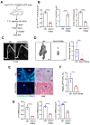
Elevated levels of Activin A in HO tissues of FOP mice. (A) Pinch injury was introduced into the right-side of the gastrocnemius muscle of the 8-week-old Acvr1R206H/+ mice, and four weeks later, trauma-induced HO was assessed via radiography (left). Alternatively, the protein levels of Activin A in tissue lysates were measured using ELISA (right, n = 3). HB: heterotopic bone. (B) Protein levels of Activin A in the supernatant of mouse Acvr1+/+;Prx1 (WT) and Acvr1R206H;Prx1 (FOP) BMSCs (n = 4). (C,D) Mouse (C, n = 4) or human (D, n = 3) BMSCs were cultured under non-differentiation (ND) or osteoblast differentiation (OBD) conditions in the presence of PBS, LPS, TNF, or IL-1β, and Activin A expression in the supernatant was measured via ELISA. (E) Mouse Acvr1R206H;Prx1 BMSCs were transduced using AAV9 carrying control (amiR-ctrl) or amiR-ACVR1R206H.ACVR1opt and then cultured under osteogenic conditions in the presence of PBS, LPS, TNF, or IL-1β. Twenty-four hours later, Activin A expression in the supernatant was measured by ELISA (n = 3). **, p < 0.01; ***, p < 0.001, and ****, p < 0.0001 by an unpaired, two-tailed Student’s t-test (AC) and one-way ANOVA test (D,E).
Figure 2
Figure 2
Generation of an Activin A-silencing AAV vector. (A) HA-tagged mouse or human Inhba plasmid was transfected into HEK293 cells along with the control (amiR-ctrl) or amiR-Inhba #1–#6 plasmid and then total cell lysates were immunoblotted with the HA antibody. Gapdh was used as a loading control. amiR-Inhba #1–#6 indicate six amiRs targeting the shared coding sequences of mouse and human Inhba. Original images can be found in Figure S3. (B) Mouse Acvr1R206H;Prx1 BMSCs or human BMSCs were transduced via AAV9 carrying amiR-ctrl or amiR-Inhba #4, and mRNA levels of Inhba were assessed using RT-PCR (n = 4). (C) AAV-transduced mouse Acvr1R206H;Prx1 BMSCs or human BMSCs were cultured in the presence of PBS, LPS, TNF, or IL-1β, and 24 h later, Activin A expression in the supernatant was measured using ELISA (n = 3). *, p < 0.05; **, p < 0.01; and ***, p < 0.001 via an unpaired two-tailed Student’s t-test (B) and one-way ANOVA test (C).
Figure 3
Figure 3
Biodistribution of AAV9.gfp.MIR in mice. A total of 5 × 10¹² vg/kg of AAV9.gfp.MIR was transdermally (t.d.) injected into 8-week-old Acvr1R206H/+ mice (n = 3), and 2 weeks later, individual tissue distribution of AAVs was assessed via EGFP expression using the IVIS optical imaging system (A) or histology on a frozen section of AAV-treated tissues (B). Scale bars: 100 μm.
Figure 4
Figure 4
AAV gene therapy suppresses Activin A signaling and osteogenic differentiation of FOP skeletal progenitors. (A) A schematic diagram showing AAV vector genome and capsid. AAV vector genome containing the CBA promoter, amiR-ACVR1R026H, amiR-Inhba #4 (red), codon-optimized human ACVR1 (ACVR1opt), and liver-abundant miR-122 target sequences (TS) was packaged into AAV9 capsid. CBA: chicken β actin; PA: poly A sequences. (BD) Mouse Acvr1R206H;Prx1 BMSCs were transduced by AAV9 carrying gfp.MIR (ctrl) or Acvr1/Inhba.MIR and cultured under osteogenic conditions. Six days later, gene expression was measured via RT-PCR and normalized to Gapdh (B,D, n = 4). Osteogenic differentiation was assessed by ALP activity (D, n = 5). (EG) AAV-transduced BMSCs were cultured under osteogenic conditions in the presence of PBS or Activin A (50 ng/mL). ALP activity (3 day culture) or alizarin red staining (10 day culture) were performed for early or late osteogenic differentiation, respectively (D, n = 5). Activin A-induced expression of Id1 and Msx2 was assessed via RT-PCR 24 h post-stimulation (E, n = 4). Cells were stimulated with Activin A at different time points and immunoblotted for phospho-Smad1/5. Hsp90 was used as a loading control (F). Original images of (G) can be found in Figure S4. **, p < 0.01; ***, p < 0.001, and ****, p < 0.0001 with an unpaired two-tailed Student’s t-test (AC) and one-way ANOVA test (D,E).
Figure 5
Figure 5
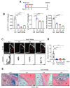
Local delivery of AAV gene therapy prevents trauma-induced HO in FOP mice. (A) Diagram of study and treatment methods. A total of 5 × 10¹² vg/kg of AAV9 carrying gfp.MIR (ctrl) or Acvr1/Inhba.MIR was injected t.d. into the gastrocnemius muscle of 8-week-old Acvr1R206H/+;Pdgfrα-GFP mice 3 days prior to pinch injury. Four weeks later, ACVR1R206H, ACVR1OPT, and Inhba expression was assessed using RT-PCR (B, n = 4), and HO in the injured muscle was assessed via X-ray (C), microCT (D), and histology on a frozen section of AAV-treated hindlimbs (E). Three-dimensional reconstruction images (D, left) and quantification of HO volume (D, right, n = 7~10) are displayed. Red arrow indicates heterotopic bone (C). Proliferation of Pdgfrα-GFP-expressing FAPs within HO tissues was visualized using fluorescence microscopy. Scale bars: 100 μm. HB: heterotopic bone; M: muscle. The frequency of GFP+ScaI+CD31CD45 FAPs in the injured muscle was assessed using flow cytometry (F), and Pdgfrα-GFP FAPs were FACS-sorted from the injured muscle and subjected to RT-PCR analysis (G, n = 4). **, p < 0.01 and ****, p < 0.0001 using an unpaired, two-tailed Student’s t-test (B,D,F,G).
Figure 6
Figure 6
Local delivery of AAV gene therapy suppresses progression of traumatic HO in FOP mice. (A) Diagram of study and treatment methods. After pinch injury on the gastrocnemius muscle of 8-week-old Acvr1R206H/+ mice at day 0, t.d. injections of AAV9.Acvr1/Inhba.MIR (5 × 10¹² vg/kg) into the injured muscle were performed on days 1, 3, or 6 post-injury. AAV9.gfp.MIR (ctrl) was t.d. injected on the same day of pinch injury. ACVR1R206H, ACVR1OPT, and Inhba expression were assessed via RT-PCR at 4 weeks post-injury (B, n = 4). HO in the injured muscle was assessed using X-ray (C, top), microCT (C, bottom), and histology on a paraffin section of AAV-treated hindlimbs (D). Three-dimensional reconstruction images (C) and quantification of HO volume (D, n = 9~10) are displayed. Red arrows indicate heterotopic bones (C). Longitudinal sections of the injured muscle was stained with Alcian blue/hematoxylin/orange G (E). HB: heterotopic bone; F: fibrotic tissue; C: chondrogenic analgen; M: skeletal muscle. Scale bars: 100 μm. ***, p < 0.001 ****, p < 0.0001 using a one-way ANOVA test (A,B,D).

Similar articles

Cited by

References

    1. Pignolo R.J., Hsiao E.C., Baujat G., Lapidus D., Sherman A., Kaplan F.S. Prevalence of fibrodysplasia ossificans progressiva (FOP) in the United States: Estimate from three treatment centers and a patient organization. Orphanet J. Rare Dis. 2021;16:350. doi: 10.1186/s13023-021-01983-2. - DOI - PMC - PubMed
    1. Pignolo R.J., Shore E.M., Kaplan F.S. Fibrodysplasia ossificans progressiva: Diagnosis, management, and therapeutic horizons. Pediatr. Endocrinol. Rev. 2013;10((Suppl. 2)):437–448. - PMC - PubMed
    1. Wentworth K.L., Masharani U., Hsiao E.C. Therapeutic advances for blocking heterotopic ossification in fibrodysplasia ossificans progressiva. Br. J. Clin. Pharmacol. 2019;85:1180–1187. doi: 10.1111/bcp.13823. - DOI - PMC - PubMed
    1. Yang Y.S., Xie J., Wang D., Kim J.M., Tai P.W.L., Gravallese E., Gao G., Shim J.H. Bone-targeting AAV-mediated silencing of Schnurri-3 prevents bone loss in osteoporosis. Nat. Commun. 2019;10:2958. doi: 10.1038/s41467-019-10809-6. - DOI - PMC - PubMed
    1. Herzog R.W. Encouraging and Unsettling Findings in Long-Term Follow-up of AAV Gene Transfer. Mol. Ther. 2020;28:341–342. doi: 10.1016/j.ymthe.2020.01.007. - DOI - PMC - PubMed

Publication types