. 2023 Sep 19;13(9):1413.
doi: 10.3390/biom13091413.

Eicosapentaenoic Acid Influences the Lipid Profile of an In Vitro Psoriatic Skin Model Produced with T Cells

Affiliations

Eicosapentaenoic Acid Influences the Lipid Profile of an In Vitro Psoriatic Skin Model Produced with T Cells

Sophie Morin et al. Biomolecules. .

Abstract

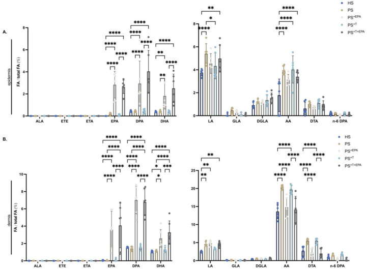
Psoriasis is a skin disease characterized by epidermal hyperplasia and an inappropriate activation of the adaptive immunity. A dysregulation of the skin's lipid mediators is reported in the disease with a predominance of the inflammatory cascade derived from n-6 polyunsaturated fatty acids (n-6 PUFAs). Bioactive lipid mediators derived from arachidonic acid (AA) are involved in the inflammatory functions of T cells in psoriasis, whereas n-3 PUFAs' derivatives are anti-inflammatory metabolites. Here, we sought to evaluate the influence of a supplementation of the culture media with eicosapentaenoic acid (EPA) on the lipid profile of a psoriatic skin model produced with polarized T cells. Healthy and psoriatic skin substitutes were produced following the auto-assembly technique. Psoriatic skin substitutes produced with or without T cells presented increased epidermal and dermal linolenic acid (LA) and AA levels. N-6 PUFA lipid mediators were strongly measured in psoriatic substitutes, namely, 13-hydroxyoctadecadienoic acid (13-HODE), prostaglandin E2 (PGE2) and 12-hydroxyeicosatetraenoic acid (12-HETE). The added EPA elevated the amounts of EPA, n-3 docosapentaenoic acid (DPA) and docosahexaenoic acid (DHA) in the epidermal and dermal phospholipids. The EPA supplementation balanced the production of epidermal lipid mediators, with an increase in prostaglandin E3 (PGE3), 12-hydroxyeicosapentaenoic acid (12-HEPE) and N-eicosapentaenoyl-ethanolamine (EPEA) levels. These findings show that EPA modulates the lipid composition of psoriatic skin substitutes by encouraging the return to a cutaneous homeostatic state.

Keywords: T cells; bioactive lipid mediators; n-3 PUFAs; psoriasis.


Conflict of interest statement

The authors declare no conflict of interest.

Figures

Figure 1
Figure 1
Histology, immunofluorescence and total phospholipid quantification of the skin substitutes. (AE) Macroscopic representations of healthy and psoriatic skin substitutes. (FJ) Histological representations of H&E staining of healthy and psoriatic skin substitutes. The scale bar represents 100 μm. (KO) Indirect immunofluorescence staining of K14 expression in healthy and psoriatic skin substitutes. K14 expression is shown in green. The cell nuclei were counterstained with DAPI reagent and are shown in blue. The dashed white lines represent the basement membrane. (P) Characterization of epidermal total fatty acids using gas chromatography. Impact of EPA supplementation on total n-3 and n-6 PUFA percentages (n-3 and n-6 PUFAs/ total fatty acids). (Q) Characterization of dermal total fatty acids using gas chromatography. Impact of EPA supplementation on total n-3 and n-6 PUFA percentages (n-3 and n-6 PUFAs/ total fatty acids). The values are presented as percentages (N = 3 donors, n = 2 skin substitutes per donor). Statistical significance was determined using two-way ANOVA followed by Tukey’s post hoc test. Significant differences are indicated by asterisks (* p < 0.05; ** p < 0.01; *** p < 0.001; **** p < 0.0001). Abbreviations: HS: healthy substitutes; EPA: eicosapentaenoic acid; PS: psoriatic substitutes; PS+EPA: psoriatic substitutes supplemented with EPA; PS+T: psoriatic substitutes produced with T cells; PS+T+EPA: psoriatic substitutes produced with T cells and supplemented with EPA; and T: T cells.
Figure 2
Figure 2
PUFA incorporation into epidermal and dermal phospholipids of the skin substitutes. Characterization of the levels of n-3 and n-6 PUFA amounts in the phospholipids of the epidermis and dermis of HS, PS, PS+EPA, PS+T and PS+T+EPA; (A) n-3 and n-6 PUFA levels in the epidermal phospholipid fraction of the skin substitutes; (B) n-3 and n-6 PUFA levels in the dermal phospholipid fraction of the skin substitutes. PUFAs were quantified using gas chromatography, and results are presented as percentages (fatty acids/total fatty acids) (N = 3 donors; n = 2 skin substitutes per donor). Statistical significance was determined using two-way ANOVA followed by Tukey’s post hoc test. Significant differences are indicated by asterisks (* p < 0.05; ** p < 0.01; *** p < 0.001; **** p < 0.0001). Abbreviations: AA: arachidonic acid; ALA: alpha-linolenic acid; DGLA: dihomo-γ-linolenic acid; DHA: docosahexaenoic acid; DPA: docosapentaenoic acid; DTA: docosatetraenoic acid; EPA: eicosapentaenoic acid; ETA: eicosatetraenoic acid; ETE: eicosatrienoic acid; GLA: γ-linolenic acid; HS: healthy substitutes; LA: linoleic acid; T: T cells; n-3: omega-3; n-6: omega-6; PUFAs: polyunsaturated fatty acids; PS: psoriatic substitutes; PS+EPA: psoriatic substitutes supplemented with EPA; PS+T: psoriatic substitutes produced with T cells; and PS+T+EPA: psoriatic substitutes produced with T cells and supplemented with EPA.
Figure 3
Figure 3
Impact of supplementation with EPA on the n-3 PUFA lipid mediator profile of the skin substitutes. Characterization of the levels of n-3-PUFA lipid mediators in the epidermis of HS, PS, PS+EPA, PS+T and PS+T+EPA. (A) Heatmap of n-3-lipid mediators analyzed using LC-MS/MS in the epidermis of the skin substitutes. (B) Characterization of epidermal n-3 prostaglandins (PG). (C) Characterization of epidermal n-3 hydroxyeicosapentaenoic acids (HEPE). (D) Characterization of epidermal n-3-hydroxydocosahexaenoic acids (HDHA) (N = 3 donors; n = 2 skin substitutes per donor). Statistical significance was determined using two-way ANOVA followed by Bonferroni’s post hoc test. Significant differences are indicated by asterisks (* p < 0.05; ** p < 0.01; *** p < 0.001; **** p < 0.0001). Abbreviations DHA: docosahexaenoic acid; EPA: eicosapentaenoic acid; HEPE: hydroxyeicosapentaenoic acid; HDHA: hydroxydocosahexaenoic acid; HS: healthy substitutes; T: T cells; n-3: omega-3; n-6: omega-6; PG: prostaglandin; PUFAs: polyunsaturated fatty acids; PS: psoriatic substitutes; PS+EPA: psoriatic substitutes supplemented with EPA; PS+T: psoriatic substitutes produced with T cells; and PS+T+EPA: psoriatic substitutes produced with T cells and supplemented with EPA.
Figure 4
Figure 4
Impact of supplementation with EPA on the n-6-PUFA lipid mediator profile of the skin substitutes. Characterization of the levels of n-6-PUFA lipid mediators in the epidermis of HS, PS, PS+EPA, PS+T and PS+T+EPA. (A) Heatmap of n-6-lipid mediators analyzed using LC-MS/MS in the epidermis of the skin substitutes. (B) Characterization of epidermal n-6 hydroxyocatadecadienoic acids (HODE). (C) Characterization of epidermal n-6 prostaglandins (PG). (D) Characterization of epidermal n-6-hydroxyeicosatetraenoic acids (HETE). Results are expressed as pmol per g of tissue. (E) Characterization of epidermal LTB4 levels (N = 3 donors; n = 2 skin substitutes per donor). Statistical significance was determined using two-way ANOVA followed by Bonferroni’s post hoc test. Significant differences are indicated by asterisks (* p < 0.05; ** p < 0.01; *** p < 0.001; **** p < 0.0001). Abbreviations: AA: arachidonic acid; HETE: hydroxyeicosatetraenoic acid; HODE: hydroxyoctadecadienoic acid; HS: healthy substitutes; LA: linoleic acid; T: T cells; n-3: omega-3; n-6: omega-6; PG: prostaglandin; PUFAs: polyunsaturated fatty acids; PS: psoriatic substitutes; PS+EPA: psoriatic substitutes supplemented with EPA; PS+T: psoriatic substitutes produced with T cells; and PS+T+EPA: psoriatic substitutes produced with T cells and supplemented with EPA.
Figure 5
Figure 5
Impact of the supplementation with EPA on the NAE profile of the skin substitutes. Characterization of the NAE derivate levels of the epidermis of HS, PS, PS+EPA, PS+T and PS+T+EPA. (A) Heatmap of the NAEs analyzed using LC-MS/MS in the epidermis of the skin substitutes. (B) Characterization of epidermal NAEs derived from n-3 PUFAs. (C) Characterization of epidermal NAEs derived from n-6 PUFAs. Results are expressed as pmol per g of tissue (N = 3 donors; n = 2 skin substitutes per donor). Statistical significance was determined using two-way ANOVA followed by Bonferroni’s post hoc test. Significant differences are indicated by asterisks (* p < 0.05; *** p < 0.001; **** p < 0.0001). Abbreviations: AEA: N-arachidoyl-ethanolamine; 13-HODE-EA: 13-hydroxyoctadecadienoic acid ethanolamine; DHEA: N-docosahexaenoyl-ethanolamine; DPEA: N-docosapentaenoyl-ethanolamine; EPEA: N-eicosapentaenoyl-ethanolamine; LEA: N-linoleoyl-ethanolamine; MUFA: monounsaturated fatty acid; NAE: N-acyl-ethanolamine; PUFAs: polyunsaturated fatty acids; PS: psoriatic substitutes; PS+EPA: psoriatic substitutes supplemented with EPA; PS+T: psoriatic substitutes produced with T cells; and PS+T+EPA: psoriatic substitutes produced with T cells and supplemented with EPA.

Similar articles

Cited by

References

    1. Takeshita J., Gelfand J.M., Li P., Pinto L., Yu X., Rao P., Viswanathan H.N., Doshi J.A. Psoriasis in the US Medicare Population: Prevalence, Treatment, and Factors Associated with Biologic Use. J. Investig. Dermatol. 2015;135:2955–2963. doi: 10.1038/jid.2015.296. - DOI - PMC - PubMed
    1. Griffiths C.E.M., Armstrong A.W., Gudjonsson J.E., Barker J. Psoriasis. Lancet. 2021;397:1301–1315. doi: 10.1016/S0140-6736(20)32549-6. - DOI - PubMed
    1. Cox A.J., Watson W. Histological variations in lesions of psoriasis. Arch. Dermatol. 1972;106:503–506. doi: 10.1001/archderm.1972.01620130035008. - DOI - PubMed
    1. Schon M.P. Adaptive and Innate Immunity in Psoriasis and Other Inflammatory Disorders. Front. Immunol. 2019;10:1764. doi: 10.3389/fimmu.2019.01764. - DOI - PMC - PubMed
    1. Leigh I.M., Navsaria H., Purkis P.E., McKay I.A., Bowden P.E., Riddle P.N. Keratins (K16 and K17) as markers of keratinocyte hyperproliferation in psoriasis in vivo and in vitro. Br. J. Dermatol. 1995;133:501–511. doi: 10.1111/j.1365-2133.1995.tb02696.x. - DOI - PubMed

Publication types

Grants and funding