Review
. 2023 Aug 31;13(9):1269.
doi: 10.3390/brainsci13091269.

The Importance of M1-and M2-Polarized Macrophages in Glioma and as Potential Treatment Targets

Affiliations
Review

The Importance of M1-and M2-Polarized Macrophages in Glioma and as Potential Treatment Targets

Jiangbin Ren et al. Brain Sci. .

Abstract

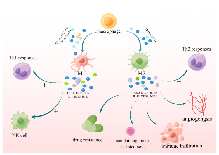
Glioma is the most common and malignant tumor of the central nervous system. Glioblastoma (GBM) is the most aggressive glioma, with a poor prognosis and no effective treatment because of its high invasiveness, metabolic rate, and heterogeneity. The tumor microenvironment (TME) contains many tumor-associated macrophages (TAMs), which play a critical role in tumor proliferation, invasion, metastasis, and angiogenesis and indirectly promote an immunosuppressive microenvironment. TAM is divided into tumor-suppressive M1-like (classic activation of macrophages) and tumor-supportive M2-like (alternatively activated macrophages) polarized cells. TAMs exhibit an M1-like phenotype in the initial stages of tumor progression, and along with the promotion of lysing tumors and the functions of T cells and NK cells, tumor growth is suppressed, and they rapidly transform into M2-like polarized macrophages, which promote tumor progression. In this review, we discuss the mechanism by which M1- and M2-polarized macrophages promote or inhibit the growth of glioblastoma and indicate the future directions for treatment.

Keywords: glioblastoma; glioma; polarization; treatment; tumor-associated macrophage.


Conflict of interest statement

The authors have no personal, financial, or institutional interests in the drugs, materials, or devices described in this article.

Figures

Figure 1
Figure 1
The polarization of macrophages and the functions of M1 and M2 TAMs. TAMs can be divided into tumor-suppressive M1-like and tumor-supportive M2-like polarized cells. M1 TAMs can secret TNF-α, IL-1β, IL-6, IL-8, IL-12, and IL-23 promoting T helper 1 (Th1) responses, and also activate natural killer (NK) cells. M2 TAMs can activate Th2-type immune response, stimulate angiogenesis, maintain tumor cell stemness, facilitate immune infiltration, and induce drug resistance by secreting ARG1, IL-13, IL-10, IL-4, VEGF, and TGF-β1. ARG-1: arginase 1; IFN-γ: interferon gamma; IL: interleukin; iNOS: inducible nitric oxide synthase; LPS: lipopolysaccharides; NK: natural killer; PPARγ: peroxisome proliferator-activated receptor-γ; STAT: signal transducer and activator of transcription 3; TAMs: tumor-associated macrophages; TGF-β: transforming growth factor-β; Th1: T helper 1; TLR2/4: Toll Like Receptor2/4; TNF-α: tumor necrosis factor; VEGF: vascular endothelial growth factor.
Figure 2
Figure 2
Partial mechanisms of M1 and M2 TAMs polarization. i: PTEN-deficient GBM cells activate PI3K/AKT pathway, which suppresses IRF1 degradation by phosphorylating glycogen synthase kinase 3 (GSK3β) into an inactive form. Then, upregulated Gal-9 interacts with Tim3 to promote the polarization of M2 TAMs. ii: Chemerin binding with CMKLR1 activates NF-κB pathway, which promotes M2 polarization. iii: JMJD1C upregulates miR-302a by promoting H3K9 demethylation at the promoter region, then negatively regulating the expression of METTL3. Therefore, downregulated METTL3 inhibits SOCS2 degradation by inhibiting m6A methylation modification to promote M1 polarization. iv: CHI3L1 regulated by the PI3K/AKT/mTOR pathway in a positive feedback loop binds with Gal3-activating NF-κB pathway to promote M2 polarization. AKT: protein kinase B; CHI3L1: chitinase-3-like protein-1; CMKLR1: chemokine-like receptor 1; Gal: galectin; Gal3BP: galectin 3-binding protein; GBM: glioblastoma; GSK3β: glycogen synthase kinase 3 beta; IRF1: interferon regulatory factor 1; JMJD1C: jumonji domain-containing 1C; NF-κB: nuclear factor kappa-B; METTL3: methyltransferase-like 3; mTOR: mammalian target of rapamycin; PIP2: phosphatidylinositol (4,5)-bisphosphate; PIP3: phosphatidylinositol (3,4,5)-triphosphate; P85: regulatory subunit; P110: catalytic subunit; PI3K: phosphatidylinositol 3 kinase; SOCS2: suppressor of cytokine signaling 2; m6A:; Tim3: T cell immunoglobulin and mucin domain 3. Meaning of symbols: +, promotion or activation; -----|, inhibition; ↑, upregulated.
Figure 3
Figure 3
Molecular mechanisms of M1 and M1 TAMs polarization. i: The deletion of MTAP often leads to the accumulation of MTA, which binds with A2B receptors to promote M2 polarization. ii: MFG-E8 binding with ITGB3 not only promotes GBM growth in an autocrine manner, but also promotes M2 polarization via activation of STAT3. iii: BACE1 acts as a transmembrane protease mediating the shedding of IL-6R, and soluble IL-6 receptor (sIL-6R) in extracellular matrix binds with IL-6 forming an IL-6/sIL-6R complex. Then, the complex binds to gp130 activating phosphorylation of STAT3 to promote M2 polarization. iv: HMGB1 activated RAGE through the phosphorylation of ERK1/2 and IKB, then activating the RAGE/NF-κB/NLRP3 inflammasome pathway, which promoted the release of TNF-α, IFN-γ, IL-1β, IL-6, IL-8, and CCL2, thereby enhancing M1-like polarization of TAMs. BACE1: β-site AβPP-cleaving enzyme 1; CDKN2A: cyclin-dependent kinase inhibitor 2A; GBM: glioblastoma; gp130: glycoprotein 130; HMGB1: high mobility group box 1 protein; ITGB3: integrin β3; MFG-E8: milk fat globule-epidermal growth factor 8; MTA: methylthioadenosine; MTAP: methylthioadenosine phosphorylase; NLRP3: NOD-like receptor thermal protein domain-associated protein 3; RAGE: receptor for advanced glycation end products; sIL-6R: soluble IL-6 receptor; STAT: signal transducer and activator of transcription 3. Meaning of symbols: +, promotion or activation; ↑, upregulated.
Figure 4
Figure 4
The treatments of the glioma. i: Binding of CD47 to SIRPα promotes phosphorylation of ITIM, which recruits and activates protein tyrosine phosphatases (PTPs). Anti-CD47 treatment not only will promote the polarization of M1 macrophages, but also promote cytotoxic phagocytosis of CD8+ T cell. ii: Anti-PD-1 treatment, α-NETA severing chemerin/CMKLR1/NF-κB axis, celastrol severing IL-13/p-STAT6 axis, anti-CD74 or anti-MIF severing MIF/CD74/p-ERK1/2/IFN-γ axis all promoted M1 polarization and inhibited M2 polarization. iii: WISP1 autocrined by GSC maintains GSCs through integrin α6β1-AKT. And it also promotes a self-renewal proliferation of M2 TAMs through a paracrine manner. α-NETA: 2-(alpha-naphthoyl)ethyltrimethylammonium; CD47: cluster of differentiation 47; CMKLR1: chemokine-like receptor 1; ERK1/2: extracellular regulated protein kinases; GSC: glioma stem cells; ITIM: immunoreceptor tyrosine-based inhibitory motifs; MHC-I: the major histocompatibility complex class I; MIF: migration inhibitory factor; PD-1: programmed cell death protein 1; PD-L1: programmed cell death 1 ligand 1; PTPs: protein tyrosine phosphatases; SIRPα: Signal Regulatory Protein α; WISP1: Wnt-induced signaling protein 1. Meaning of symbols: +, promotion or activation; -----|, inhibition.

Similar articles

Cited by

References

    1. Codrici E., Popescu I.D., Tanase C., Enciu A.M. Friends with Benefits: Chemokines, Glioblastoma-Associated Microglia/Macrophages, and Tumor Microenvironment. Int. J. Mol. Sci. 2022;23:2509. doi: 10.3390/ijms23052509. - DOI - PMC - PubMed
    1. Wang L.J., Lv P., Lou Y. Alarm Signal S100-Related Signature Is Correlated with Tumor Microenvironment and Predicts Prognosis in Glioma. Dis. Markers. 2022;2022:4968555. doi: 10.1155/2022/4968555. - DOI - PMC - PubMed
    1. Seyfrid M., Maich W.T., Shaikh V.M., Tatari N., Upreti D., Piyasena D., Subapanditha M., Savage N., McKenna D., Mikolajewicz N., et al. CD70 as an actionable immunotherapeutic target in recurrent glioblastoma and its microenvironment. J. Immunother. Cancer. 2022;10:e003289. doi: 10.1136/jitc-2021-003289. - DOI - PMC - PubMed
    1. Shi Y., Ping Y.F., Zhou W., He Z.C., Chen C., Bian B.S., Zhang L., Chen L., Lan X., Zhang X.C., et al. Tumour-associated macrophages secrete pleiotrophin to promote PTPRZ1 signalling in glioblastoma stem cells for tumour growth. Nat. Commun. 2017;8:15080. doi: 10.1038/ncomms15080. - DOI - PMC - PubMed
    1. Pancholi S., Tripathi A., Bhan A., Acharya M.M., Pillai P. Emerging Concepts on the Role of Extracellular Vesicles and Its Cargo Contents in Glioblastoma-Microglial Crosstalk. Mol. Neurobiol. 2022;59:2822–2837. doi: 10.1007/s12035-022-02752-3. - DOI - PMC - PubMed

LinkOut - more resources