. 2023 Sep 8;13(9):1298.
doi: 10.3390/brainsci13091298.

Repetitive, but Not Single, Mild Blast TBI Causes Persistent Neurological Impairments and Selective Cortical Neuronal Loss in Rats

Affiliations

Repetitive, but Not Single, Mild Blast TBI Causes Persistent Neurological Impairments and Selective Cortical Neuronal Loss in Rats

Rita Campos-Pires et al. Brain Sci. .

Abstract

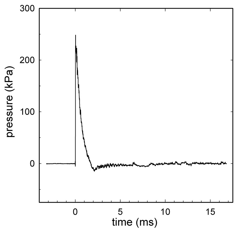
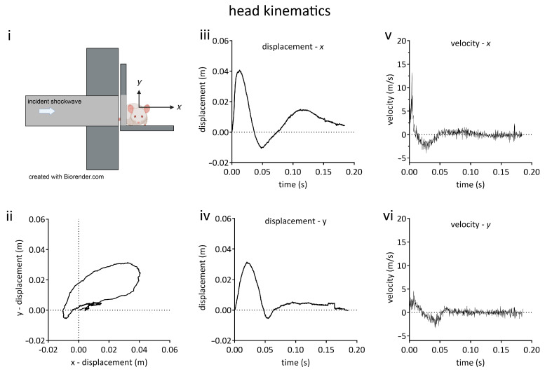
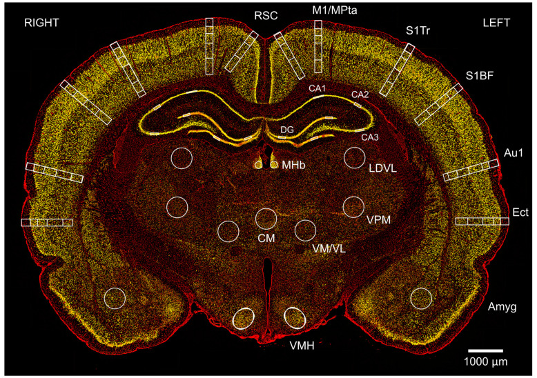
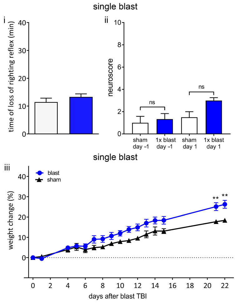
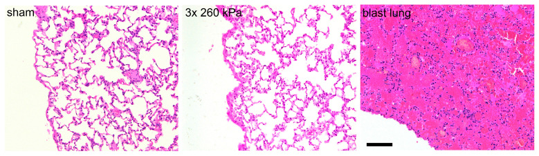
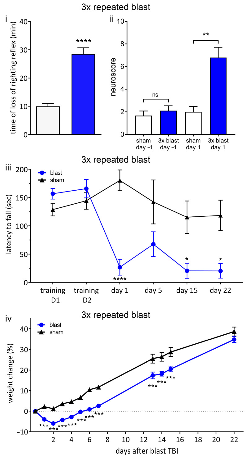
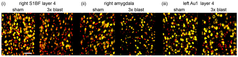
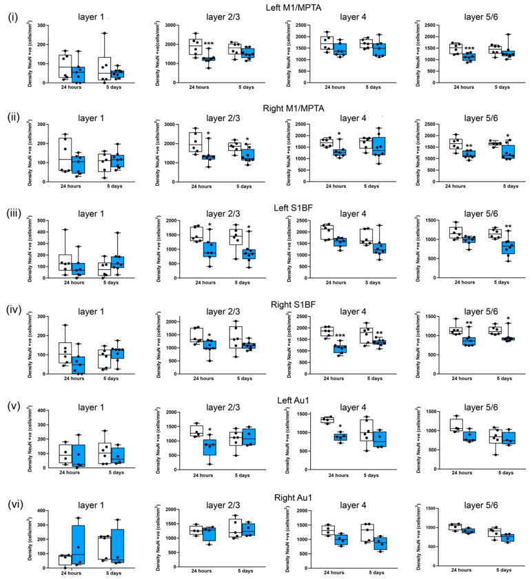
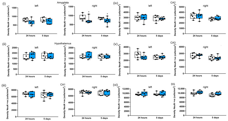
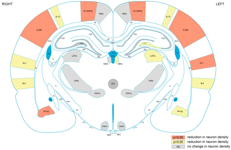
Exposure to repeated mild blast traumatic brain injury (mbTBI) is common in combat soldiers and the training of Special Forces. Evidence suggests that repeated exposure to a mild or subthreshold blast can cause serious and long-lasting impairments, but the mechanisms causing these symptoms are unclear. In this study, we characterise the effects of single and tightly coupled repeated mbTBI in Sprague-Dawley rats exposed to shockwaves generated using a shock tube. The primary outcomes are functional neurologic function (unconsciousness, neuroscore, weight loss, and RotaRod performance) and neuronal density in brain regions associated with sensorimotor function. Exposure to a single shockwave does not result in functional impairments or histologic injury, which is consistent with a mild or subthreshold injury. In contrast, exposure to three tightly coupled shockwaves results in unconsciousness, along with persistent neurologic impairments. Significant neuronal loss following repeated blast was observed in the motor cortex, somatosensory cortex, auditory cortex, and amygdala. Neuronal loss was not accompanied by changes in astrocyte reactivity. Our study identifies specific brain regions particularly sensitive to repeated mbTBI. The reasons for this sensitivity may include exposure to less attenuated shockwaves or proximity to tissue density transitions, and this merits further investigation. Our novel model will be useful in elucidating the mechanisms of sensitisation to injury, the temporal window of sensitivity and the evaluation of new treatments.

Keywords: blast neurotrauma; blast trauma; blast traumatic brain injury; concussion; functional deficits; repetitive brain injury.


Conflict of interest statement

Dickinson received funding as detailed above and acknowledges the financial support of the Royal British Legion. Rita Campos-Pires was the recipient of a doctoral training award from the Fundação para a Ciência e a Tecnologia, Lisbon, Portugal, and received funding from the Association of Paediatric Anaesthetists of Great Britain and Ireland. Mariia Koziakova was the recipient of a Rector’s PhD Studentship from Imperial College London. Eszter Ujvari is the recipient of an MRC PhD studentship. The other authors declare no competing interests.

Figures

Figure 1
Figure 1
A representative shock wave recorded during the experiment with animal in position. A signal obtained from a radial pressure sensor (2300 V1, Dytran Instruments, Chatsworth, CA, USA) at the distal end of the shock tube: the radial peak overpressure was 246 kPa; the positive wave duration was 1.3 ms; the impulse was 117 kPa·ms. Compressed air was used in the shock tube with a single diaphragm configuration using 225 µm Mylar®. Data were recorded using a high-bandwidth oscilloscope (Tektronix model DPO4104B Tektronix Inc., Beaverton, OR, USA) at a sampling rate of 50 MHz, before being digitally filtered offline at 40 kHz.
Figure 2
Figure 2
Kinematic characterisation of rat head movement in the blast neurotrauma model. (i) Schematic diagram of animal orientation and positioning on platform at distal end of shock tube. Rats were oriented with the right side of the head towards incident shockwave, being positioned such that only the head was exposed and the thorax was shielded from the shockwave. The head could move unrestrained in the x-y plane. (ii) Head displacement in x-y plane. (iii) Head x-displacement as a function of time. (iv) Head y-displacement as a function of time. (v) Velocity in x axis as a function of time. (vi) Velocity in y axis as a function of time. Lines shown are the means of data obtained from 3 animals.
Figure 3
Figure 3
Changes in neuronal density after single and repeated blast injury were mapped in cortical and subcortical regions in coronal brain sections. Image shows a typical section at Bregma −3.36 mm from a sham animal at 24 h, stained with neuronal marker NeuN (yellow) and non-specific nuclear marker DAPI (red). Neurons were counted in left and right retrosplenial cortex (RSC) in layers 1, 2/3/4, and 5/6; the motor/medial parietal association cortex (M1/MPtA) was assessed in layers 1, 2/3, 4, and 5/6; the somatosensory cortex trunk region and barrel field (S1Tr, S1BF) were assessed in layers 1, 2/3, 4, and 5/6; the auditory cortex (Au1) was assessed in layers 1, 2/3, 4, and 5/6; and the ectorhinal cortex (Ect) was assessed in layers 1, 2/3, 4, and 5/6. Finally, in subcortical regions of amygdala (Amyg), ventromedial hypothalamus (VMH), centromedial thalamic nucleus (CM), ventromedial/ventrolateral thalamic nucleus (VM/VL), ventral posteromedial thalamic nucleus (VPM), laterodorsal thalamic nucleus, ventrolateral (LDVL), and medial habenular nucleus (MHb) and hippocampus (CA1; CA2; CA3 and DG) were assessed. The scale bar is 1000 μm.
Figure 4
Figure 4
Single blast does not result in unconsciousness or persistent neurological deficits. (i) A single 260-kilopascal shockwave does not result in blast-associated unconsciousness. Time until recovery of righting reflex immediately after blast (blue bar) was not significantly different to that of sham procedure (white bar), indicating that there was no blast-associated unconsciousness. (ii) Neurological outcome score in blast and sham groups was not significantly different one day before blast or one day after blast. (iii) Weight change relative to procedure day up to 22 days following blast or sham procedure. There was no weight loss in either sham or blast group. Values shown are means, and error bars are standard errors. ** p < 0.01, ns not significant. Loss of righting reflex, 1 × blast n = 22, sham n = 18, Mann–Whitney test. Neuroscore 1 × blast n = 6, sham n = 4, Kruskal–Wallis test with Benjamini–Yekutieli correction. Weight change, 1 × blast n = 16, sham n = 10, two-way ANOVA with Sidak’s correction.
Figure 5
Figure 5
Head-only blast model avoids lung injury. Typical haematoxylin- and eosin-stained slices obtained from right caudal lobe of lungs of animals exposed to sham procedure or three repeated shockwaves at 24 h after the blast or sham procedure. Also shown is right caudal lobe obtained from an animal with fatal blast lung. The alveolar spaces in the sham and three repeated blast animals are clear, well inflated, and free of red blood cells. In contrast, the tissue obtained from animal with blast lung exhibits severe pulmonary haemorrhage, or ‘hepatisation’. Scale bar is 100 μm.
Figure 6
Figure 6
Repeated blast results in unconsciousness and persistent neurological deficits. (i) Three repeated shockwaves result in blast-associated unconsciousness. Time until recovery of righting reflex immediately after blast (blue bar) was significantly increased compared to sham procedure (white bar), indicating blast-induced unconsciousness. (ii) There are neurological impairments after three repeated shockwaves. One day before procedure, there was no significant difference between blast (blue bar) and sham (white bar) groups. Following blast, there was a significant increase in neuroscore in blast group compared to sham group, indicating neurological impairments. (iii) RotaRod performance indicates persistent vestibulomotor deficits following repeated blast exposure. There was no difference in latency to fall between blast and sham groups on two training days before injury. Latency to fall was significantly decreased in blast group on day 1, day 15, and day 22 after blast. (iv) Weight change relative to procedure day up to 22 days following blast or sham procedure. There was no weight loss in sham group. There was significant weight loss in repeated blast group compared to sham group. Values shown are means, and error bars are standard errors. * p < 0.05, ** p < 0.01 *** p < 0.001, **** p < 0.0001, ns not significant. Loss of righting reflex, 3 × blast n = 28, n = 22 sham, Mann–Whitney test. Neuroscore 3 × blast n = 10, sham n = 9, Kruskal–Wallis test with Benjamini–Yekutieli correction. Weight change, 3 × blast n = 25, sham, n = 22, two-way ANOVA with Sidak’s correction. Rotarod; 3 × blast n = 15, sham n = 12, two-way ANOVA with Sidak’s correction.
Figure 7
Figure 7
Repeated 260-kilopascal blast results in neuronal loss. Representative neuronal staining in sham and three repeated blast groups in (i) right somatosensory cortex (S1BF) layer 4, (ii) right amygdala, and (iii) left auditory cortex (Au1) layer 4. NeuN positive cells are shown in yellow, and DAPI is shown in red. Images represent areas included in neuronal quantification in Figure 7 and Figure 8. The scale bar is 50 μm.
Figure 8
Figure 8
Three repeated shockwaves result in cortical neuronal loss. Quantification of neuronal cell density of cortical layers from sham (white boxes) and three repeated 260-kilopascal blasts (blue boxes) groups in (i) left and (ii) right motor/medial parietal association cortex (M1/MPtA), (iii) left and (iv) right somatosensory cortex (S1BF), and (v) left and (vi) right auditory cortex (Au1). Lines are medians, boxes represent interquartile intervals, and whiskers are ranges. * p < 0.05, ** p < 0.01, and *** p < 0.001 compared to sham; Kruskal–Wallis test with Benjamini–Yekutieli correction. After 24 h, sham n = 6 and blast n = 7; after 5 days, sham n = 6, and, blast n = 8. Not all slices included Au1.
Figure 9
Figure 9
Three repeated shockwaves of 260 kPa result in selective subcortical neuronal loss. Quantification of neuronal cell density in left and right (i) amygdala; (ii) hypothalamus and hippocampal (iii) CA1, (iv) CA2, (v) CA3, and (vi) DG subregions; and in sham (white boxes) in repeated 260-kilopascal blasts (blue boxes). The lines are medians, boxes are interquartile intervals, and whiskers are ranges. * p < 0.05 compared to sham; Kruskal–Wallis test with Benjamini–Yekutieli correction. After 24 h, sham n = 6 and blast n = 7; after 5 days, sham n = 6 and blast n = 8.
Figure 10
Figure 10
Cortical brain areas are most sensitive to repeated blast TBI. Mapping of regions of interest analysed exhibiting significant (p < 0.05) reductions neuronal density (orange), reductions in neuronal density that did not reach significance (yellow), or no change in neuronal density (grey). RSC, retrosplenial cortex; M1/MPtA, motor cortex/medial parietal temporal area; S1Tr, somatosensory cortex trunk area; S1BF, somatosensory cortex, barrel field; Au1, primary auditory cortex; Ect, ectorhinal cortex; Amyg, amygdala; VMH, ventromedial hypothalamic nucleus; VM/VL, ventromedial/ventrolateral thalamic nucleus; VPM, ventral posteromedial thalamic nucleus; LDVL, laterodorsal thalamic nucleus, ventrolateral; MHb, medial habenular nucleus; CA1, hippocampal CA1 region: CA2, hippocampal CA2 region; CA3, hippocampal CA3 region; DG, hippocampal dentate gyrus region. The ventricles are shown in blue. Figure 10 is based on rat brain atlas of Paxinos and Watson [74], and is used with permission.

Similar articles

Cited by

References

    1. Taylor C.A., Bell J.M., Breiding M.J., Xu L. Traumatic Brain Injury-Related Emergency Department Visits, Hospitalizations, and Deaths-United States, 2007 and 2013. MMWR Surveill. Summ. 2017;66:1–16. doi: 10.15585/mmwr.ss6609a1. - DOI - PMC - PubMed
    1. Maas A.I.R., Menon D.K., Adelson P.D., Andelic N., Bell M.J., Belli A., Bragge P., Brazinova A., Buki A., Chesnut R.M., et al. Traumatic brain injury: Integrated approaches to improve prevention, clinical care, and research. Lancet Neurol. 2017;16:987–1048. doi: 10.1016/S1474-4422(17)30371-X. - DOI - PubMed
    1. Wojcik B.E., Stein C.R., Bagg K., Humphrey R.J., Orosco J. Traumatic brain injury hospitalizations of U.S. army soldiers deployed to Afghanistan and Iraq. Am. J. Prev. Med. 2010;38((Suppl. S1)):S108–S116. doi: 10.1016/j.amepre.2009.10.006. - DOI - PubMed
    1. Chapman J.C., Diaz-Arrastia R. Military traumatic brain injury: A review. Alzheimers Dement. 2014;10((Suppl. S3)):S97–S104. doi: 10.1016/j.jalz.2014.04.012. - DOI - PubMed
    1. DoD_Worldwide_Numbers_for_TBI. [(accessed on 31 July 2023)]. Available online: https://dvbic.dcoe.mil/dod-worldwide-numbers-tbi.