Relation of MRI-Visible Perivascular Spaces and Other MRI Markers of Cerebral Small Vessel Disease
- PMID: 37759924
- PMCID: PMC10527297
- DOI: 10.3390/brainsci13091323
Relation of MRI-Visible Perivascular Spaces and Other MRI Markers of Cerebral Small Vessel Disease
Abstract
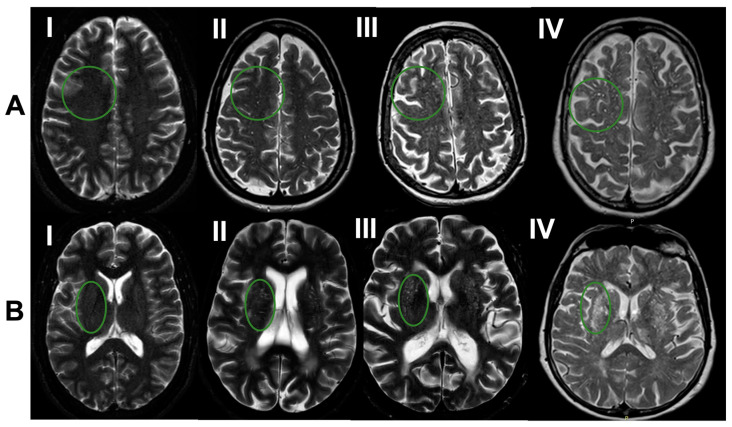
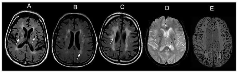
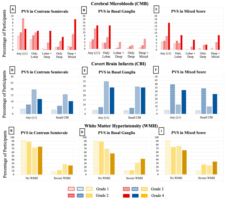
Perivascular spaces (PVS) visible on brain MRI signal cerebral small vessel disease (CSVD). The coexistence of PVS with other CSVD manifestations likely increases the risk of adverse neurological outcomes. We related PVS to other CSVD manifestations and brain volumes that are markers of vascular brain injury and neurodegeneration. Framingham Heart Study (FHS) participants with CSVD ratings on brain MRI were included. PVS were rated in the basal ganglia (BG) and centrum semiovale (CSO) into grades I-IV and a category reflecting high burden in single or mixed CSO-BG regions. We related PVS to covert brain infarcts (CBI), white matter hyperintensities (WMH), cerebral microbleeds (CMB), total brain, hippocampal, and cortical gray matter volumes using adjusted multivariable regression analyses. In 2454 participants (mean age 54 ± 12 years), we observed that higher PVS burden in both BG and CSO was related to CMB in lobar and deep brain regions and increased WMH. Greater CSO PVS burden was associated with decreased total cortical gray volumes. PVS are associated with ischemic markers of CSVD and neurodegeneration markers. Further studies should elucidate the causality between PVS and other CSVD manifestations.
Keywords: MRI-visible perivascular spaces; cerebral small vessel disease; disease marker; glymphatic function.
Conflict of interest statement
The authors declare no conflict of interest.
Figures


Similar articles
-
Relation of MRI visible perivascular spaces with global and regional brain structural connectivity measures: The Framingham Heart Study (FHS).Neurobiol Aging. 2025 Jun;150:1-8. doi: 10.1016/j.neurobiolaging.2025.02.007. Epub 2025 Feb 27. Neurobiol Aging. 2025. PMID: 40043467
-
Free water as a potential mediator linking basal ganglia peri-vascular spaces to white matter hyperintensities in cerebral small vessel disease.Front Neurosci. 2025 Jul 3;19:1621023. doi: 10.3389/fnins.2025.1621023. eCollection 2025. Front Neurosci. 2025. PMID: 40678759 Free PMC article.
-
Cerebral small vessel disease in memory center patients with dementia with lewy bodies and Alzheimer's disease.Alzheimers Res Ther. 2025 Jul 12;17(1):154. doi: 10.1186/s13195-025-01805-8. Alzheimers Res Ther. 2025. PMID: 40646592 Free PMC article.
-
The global burden of cerebral small vessel disease in low- and middle-income countries: A systematic review and meta-analysis.Int J Stroke. 2023 Jan;18(1):15-27. doi: 10.1177/17474930221137019. Epub 2022 Nov 25. Int J Stroke. 2023. PMID: 36282189
-
Cerebral small vessel disease and prognosis in intracerebral haemorrhage: A systematic review and meta-analysis of cohort studies.Eur J Neurol. 2022 Aug;29(8):2511-2525. doi: 10.1111/ene.15363. Epub 2022 May 1. Eur J Neurol. 2022. PMID: 35435301
Cited by
-
Imaging-validated correlates and implications of the pathophysiologic mechanisms of ageing-related cerebral large artery and small vessel diseases: a systematic review and meta-analysis.Behav Brain Funct. 2025 Apr 22;21(1):12. doi: 10.1186/s12993-025-00274-1. Behav Brain Funct. 2025. PMID: 40264233 Free PMC article.
-
Imaging the neurovascular unit in health and neurodegeneration: a scoping review of interdependencies between MRI measures.Fluids Barriers CNS. 2023 Dec 21;20(1):97. doi: 10.1186/s12987-023-00499-0. Fluids Barriers CNS. 2023. PMID: 38129925 Free PMC article.
-
Relation of MRI visible perivascular spaces with global and regional brain structural connectivity measures: The Framingham Heart Study (FHS).Neurobiol Aging. 2025 Jun;150:1-8. doi: 10.1016/j.neurobiolaging.2025.02.007. Epub 2025 Feb 27. Neurobiol Aging. 2025. PMID: 40043467
-
Enlarged perivascular spaces are associated with white matter injury, cognition and inflammation in cerebral autosomal dominant arteriopathy with subcortical infarcts and leukoencephalopathy.Brain Commun. 2024 Mar 8;6(2):fcae071. doi: 10.1093/braincomms/fcae071. eCollection 2024. Brain Commun. 2024. PMID: 38495305 Free PMC article.
-
Impact of enlarged perivascular spaces in the basal ganglia on gait in cerebral small vessel disease.Aging Clin Exp Res. 2025 Apr 30;37(1):138. doi: 10.1007/s40520-025-03045-0. Aging Clin Exp Res. 2025. PMID: 40304928 Free PMC article.
References
Grants and funding
- R01 NS017950/NS/NINDS NIH HHS/United States
- R01 AG054076/AG/NIA NIH HHS/United States
- R01 AG059725/AG/NIA NIH HHS/United States
- R21 NS135268/NS/NINDS NIH HHS/United States
- R01 AG059725; AG008122; AG054076; K23AG038444; R03 AG048180-01A1; AG033193/AG/NIA NIH HHS/United States
- R01 AG008122/AG/NIA NIH HHS/United States
- R01-NS017950-37/NS/NINDS NIH HHS/United States
- R01 AG080821/AG/NIA NIH HHS/United States
- R03 AG048180/AG/NIA NIH HHS/United States
- P30 AG072972/AG/NIA NIH HHS/United States
- N01 HC025195/HL/NHLBI NIH HHS/United States
- R01 AG033193/AG/NIA NIH HHS/United States
- K23 AG038444/AG/NIA NIH HHS/United States
- P30 AG010129/AG/NIA NIH HHS/United States
LinkOut - more resources
Full Text Sources
Research Materials