Review
. 2023 Aug 28;12(9):1680.
doi: 10.3390/antiox12091680.

Thioredoxin/Glutaredoxin Systems and Gut Microbiota in NAFLD: Interplay, Mechanism, and Therapeutical Potential

Affiliations
Review

Thioredoxin/Glutaredoxin Systems and Gut Microbiota in NAFLD: Interplay, Mechanism, and Therapeutical Potential

Minghui Zhu et al. Antioxidants (Basel). .

Abstract

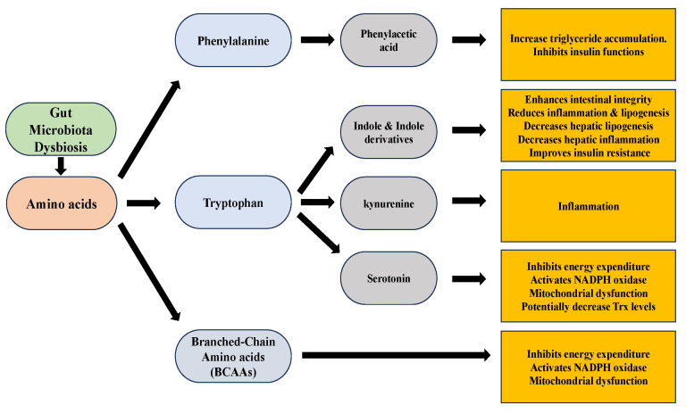
Non-alcoholic fatty liver disease (NAFLD) is a common clinical disease, and its pathogenesis is closely linked to oxidative stress and gut microbiota dysbiosis. Recently accumulating evidence indicates that the thioredoxin and glutaredoxin systems, the two thiol-redox dependent antioxidant systems, are the key players in the NAFLD's development and progression. However, the effects of gut microbiota dysbiosis on the liver thiol-redox systems are not well clarified. This review explores the role and mechanisms of oxidative stress induced by bacteria in NAFLD while emphasizing the crucial interplay between gut microbiota dysbiosis and Trx mediated-redox regulation. The paper explores how dysbiosis affects the production of specific gut microbiota metabolites, such as trimethylamine N-oxide (TMAO), lipopolysaccharides (LPS), short-chain fatty acids (SCFAs), amino acids, bile acid, and alcohol. These metabolites, in turn, significantly impact liver inflammation, lipid metabolism, insulin resistance, and cellular damage through thiol-dependent redox signaling. It suggests that comprehensive approaches targeting both gut microbiota dysbiosis and the thiol-redox antioxidant system are essential for effectively preventing and treating NAFLD. Overall, comprehending the intricate relationship between gut microbiota dysbiosis and thiol-redox systems in NAFLD holds significant promise in enhancing patient outcomes and fostering the development of innovative therapeutic interventions.

Keywords: NAFLD; gut microbiota dysbiosis; oxidative stress; reactive oxygen species; thioredoxin.


Conflict of interest statement

The authors declare no conflict of interest.

Figures

Figure 1
Figure 1
The thioredoxin system plays a crucial role in modulating cell viability and proliferation. Thioredoxin can donate electrons to various enzymes, including peroxiredoxins, which have critical roles in cell signaling by either removing hydrogen peroxide or regulating redox-sensitive signaling molecules [40,41,42,43,44,45]. The redox state of thioredoxin can affect the function of several transcription factors, making it an important player in cellular signaling [46,47,48,49]. (ER: Endoplasmic reticulum, Keap1: kelch like ECH associated protein 1, Nrf2: Nuclear factor erythroid 2-related factor 2, ARE: Antioxidant response element, TrxR: Thioredoxin Reductase, Trx: Thioredoxin, Prx: Peroxiredoxin, NADPH: Nicotinamide Adenine Dinucleotide Phosphate).
Figure 2
Figure 2
The GSH-Grx 1 system controls protein S-glutathionylation major players in the NAFLD. (GR: Glutathione Reductase, GSH: Glutathione, NADPH: Nicotinamide Adenine Dinucleotide Phosphate, Grx: Glutaredoxin, SirT-1: sirtuin type 1, PPAR: Peroxisome proliferators-activated receptors, PGC-1α: Peroxisome proliferator-activated receptor-gamma coactivator, SREBP-1c: Sterol regulatory element binding protein-1c).
Figure 3
Figure 3
Thioredoxin and glutathione system participants in defense against oxidative stress caused by gut microbiota dysbiosis in NAFLD. This figure illustrates the dynamic interplay among gut microbiota dysbiosis, oxidative stress, mitochondrial dysfunction, endoplasmic reticulum stress, inflammation, and peroxisome-related processes in the pathogenesis of NAFLD. (LPS: lipopolysaccharides, TMAO: trimethylamine N-oxide, AAs: Amino acids, DCA: deoxycholic acid, NF-κB: Nuclear Factor-kappa B, Nrf2: Nuclear factor erythroid 2-related factor 2, ERO1: Endoplasmic Reticulum Oxidoreductin 1, NOX: NADPH Oxidase, GR: Glutathione Reductase, GSH: Glutathione, TrxR: Thioredoxin Reductase, Trx: Thioredoxin, NADPH: Nicotinamide Adenine Dinucleotide Phosphate, Grx1: Glutaredoxin 1, GPx1: Glutathione Peroxidase 1, Prx1: Peroxiredoxin 1, Prx2: Peroxiredoxin 2).
Figure 4
Figure 4
Trx regulation in TMAO-caused inflammation. TMAO induces the activation of FoxO1 in the liver by binding to PERK, an ER stress sensor, which initiates the unfolded protein response (UPR). This process not only triggers oxidative stress but also contributes to the development of metabolic syndrome. This, in turn, triggers the NLRP3 inflammasome through the NF-κB pathway. This activation of NF-κB may stem from oxidant release due to ER-mediated stress, leading to the dissociation of TXNIP from Trx and subsequent binding and activation of NF-κB. NF-κB then prompts the induction of NLRP3, leading to the assembly with ASC and procaspase-1. This leads to caspase-1-mediated conversion of pro-IL-1β to the activated form IL-1β and triggering the inflammation. TMAO exposure causes oxidative stress and inflammatory cytokine release in endothelial cells, contributing to metabolic disease. (TMAO: trimethylamine N-oxide, PERK: PKR-like eukaryotic initiation factor 2α kinase, UPR: Unfolded Protein Response, NLRP3: NOD-like receptor protein 3, NF-κB: Nuclear Factor-Kappa B, TXNIP: Thioredoxin-Interacting Protein, ASC: Apoptosis-associated speck-like protein containing a CARD).
Figure 5
Figure 5
Role of Trx system in LPS impact on liver cells. LPS initiates inflammation by activating TLR4 on various cells. In the liver, LPS binding to TLR4 in macrophages (Kupffer cells) leads to the release of TNF-α and IL-6, affecting HSCs. Concurrently, iNOS induces the generation of oxidants in Kupffer cells, while activated HSCs produce oxidants through NADPH oxidase, resulting in oxidative stress and inflammation. The antioxidant Trx counteracts LPS-induced apoptosis by inhibiting the activation of ASK1, with Trx-2 located in mitochondria, regulating apoptosis signaling and collectively protecting against cell death from oxidants. Additionally, Trx inhibits HSC proliferation, reduces fibrosis, and interacts with inflammatory pathways, offering protective effects. Trx-2 overexpression reduces inflammation, neutrophil infiltration, and liver injury caused by LPS exposure, contributing to hepatic health. (LPS: Lipopolysaccharide, TLR4: Toll-like receptor 4, TNF-α: Tumor Necrosis Factor-Alpha, IL-6: Interleukin-6, iNOS: Inducible Nitric Oxide Synthase, NADPH: Nicotinamide Adenine Dinucleotide Phosphate, ASK1: Apoptosis Signal-Regulating Kinase 1, Trx: Thioredoxin, Trx-2: Mitochondrial Thioredoxin)”.
Figure 6
Figure 6
Amino acids metabolites and their effects.
Figure 7
Figure 7
Impact of DCA on the hepatocytes and antioxidant system. Elevated concentrations of DCA, acting as an antagonist of FXR in NAFLD, have the potential to antagonize FXR signaling. This antagonistic effect may lead to the activation of NF-κB, a transcription factor central to inflammation. This intriguing interaction could potentially influence the function of the thioredoxin antioxidant system. (DCA: Deoxycholic Acid, FXR: Farnesoid X Receptor, NF-κB: Nuclear Factor-Kappa B).

Similar articles

Cited by

References

    1. Stefan N., Häring H.U., Cusi K. Non-alcoholic fatty liver disease: Causes, diagnosis, cardiometabolic consequences, and treatment strategies. Lancet. Diabetes Endocrinol. 2019;7:313–324. doi: 10.1016/S2213-8587(18)30154-2. - DOI - PubMed
    1. Friedman S.L., Neuschwander-Tetri B.A., Rinella M., Sanyal A.J. Mechanisms of NAFLD development and therapeutic strategies. Nat. Med. 2018;24:908–922. doi: 10.1038/s41591-018-0104-9. - DOI - PMC - PubMed
    1. Eslam M., Sanyal A.J., George J. MAFLD: A Consensus-Driven Proposed Nomenclature for Metabolic Associated Fatty Liver Disease. Gastroenterology. 2020;158:1999–2014. doi: 10.1053/j.gastro.2019.11.312. - DOI - PubMed
    1. Seghieri M., Christensen A.S., Andersen A., Solini A., Knop F.K., Vilsbøll T. Future Perspectives on GLP-1 Receptor Agonists and GLP-1/glucagon Receptor Co-agonists in the Treatment of NAFLD. Front. Endocrinol. 2018;9:649. doi: 10.3389/fendo.2018.00649. - DOI - PMC - PubMed
    1. Buzzetti E., Pinzani M., Tsochatzis E.A. The multiple-hit pathogenesis of non-alcoholic fatty liver disease (NAFLD) Metab. Clin. Exp. 2016;65:1038–1048. doi: 10.1016/j.metabol.2015.12.012. - DOI - PubMed

LinkOut - more resources