. 2023 Aug 29;12(9):1683.
doi: 10.3390/antiox12091683.

Pharmacologic Ascorbate and DNMT Inhibitors Increase DUOX Expression and Peroxide-Mediated Toxicity in Pancreatic Cancer

Affiliations

Pharmacologic Ascorbate and DNMT Inhibitors Increase DUOX Expression and Peroxide-Mediated Toxicity in Pancreatic Cancer

Garett J Steers et al. Antioxidants (Basel). .

Abstract

Recent studies have demonstrated an important role for vitamin C in the epigenetic regulation of cancer-related genes via DNA demethylation by the ten-eleven translocation (TET) methylcytosine dioxygenase enzymes. DNA methyltransferase (DNMT) reverses this, increasing DNA methylation and decreasing gene expression. Dual oxidase (DUOX) enzymes produce hydrogen peroxide (H2O2) in normal pancreatic tissue but are silenced in pancreatic cancer (PDAC). Treatment of PDAC with pharmacologic ascorbate (P-AscH-, intravenous, high dose vitamin C) increases DUOX expression. We hypothesized that inhibiting DNMT may act synergistically with P-AscH- to further increase DUOX expression and cytotoxicity of PDAC. PDAC cells demonstrated dose-dependent increases in DUOX mRNA and protein expression when treated with DNMT inhibitors. PDAC cells treated with P-AscH- + DNMT inhibitors demonstrated increased DUOX expression, increased intracellular oxidation, and increased cytotoxicity in vitro and in vivo compared to either treatment alone. These findings suggest a potential therapeutic, epigenetic mechanism to treat PDAC.

Keywords: DNA methyltransferase (DNMT); ascorbic acid; epigenetics; pancreatic cancer; pharmacologic ascorbate; ten-eleven translocation (TET) methylcytosine dioxygenase.


Conflict of interest statement

The authors declare no conflict of interest.

Figures
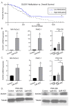
Figure 1
Figure 1
DUOX hypermethylation in PDAC. (A) DUOX1 methylation vs. overall survival in PDAC. The NIH Cancer Genome Atlas (TCGA) Pancreatic Cancer Database (PAAD) (n = 223) was accessed via the University of California Santa Cruz (UCSC) Xena Functional Genomics Explorer (n = 148; overall survival at 5 years 30% vs. 5%; * p = 0.03; Gehan–Breslow–Wilcoxon test). (B) DUOX1 mRNA expression is increased in a dose-dependent manner in the MIA PaCa-2, PANC-1, and PDX-339 PDAC cell lines after exposure to AZC (1–2 µM) for 5 days (means ± SEM; n = 3; * p < 0.05). (C) DUOX2 mRNA expression is increased in a dose-dependent manner in MIA PaCa-2, PANC-1, and PDX-339 PDAC cell lines after exposure to AZC (1–2 µM) for 5 days (means ± SEM; n = 3; * p < 0.05). (D) DUOX1 and DUOX2 immunoreactive protein was increased in PDX-339 cells after AZC (1–2 µM). Representative blots are shown.
Figure 2
Figure 2
DNMT inhibitors with P-AscH increase DUOX expression in PDAC cell lines in a dose-dependent manner. Treatment with a DNMT inhibitor also produces sustained increases in DUOX expression. (A) DUOX1 mRNA expression is increased after exposure to AZC (2 µM) for 5 days ± P-AscH (20 pmol/cell) for 1 h in PDX-339 cells. The combination group demonstrated a significant increase in expression compared to either treatment group alone (means ± SEM; n = 3; * p < 0.05). (B) DUOX1 mRNA expression increased in a dose-dependent manner after exposure to AZD (0.5–1 µM) for 3 days in MIA PaCa-2 cells. The addition of P-AscH (10 pmol/cell) for 1 h produces a significant increase in expression compared to either treatment group alone (means ± SEM; n = 3; * p < 0.05). (C) DUOX2 mRNA expression increased in a similar manner with the same treatments. (D) DUOX1 immunoreactive protein increased in MIA-PaCa-2 cells with AZD (0.5–1 µM) for 3 days and P-AscH (10 pmol/cell) for 1 h. (E) DUOX2 immunoreactive protein increased with the same treatments. Representative blots are shown. (F) DUOX1 mRNA expression is increased for up to 72 h after exposure to AZD (1 µM). (G) DUOX2 mRNA expression is increased immediately following exposure to AZD (1 µM) (means ± SEM; n = 4; * p < 0.05 vs. control).
Figure 3
Figure 3
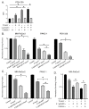
DNMT inhibitors and P-AscH generate dose-dependent, H2O2-dependent cytotoxicity. (A) Mean fluorescence intensity (MFI) is increased following exposure to AZC (2 µM) for 5 days and P-AscH (20 pmol/cell) for 1 h in PDX-339 cells. Pretreatment with bovine catalase (100 µg/mL) reverses this effect demonstrating that the oxidation of DCFH-DA is mediated by H2O2 (means ± SEM; n = 3; * p < 0.05). (B) MIA PaCa-2, PANC-1 and PDX-339 cells were treated with AZC (0.5–2 µM) for 5 days ± P-AscH (20 pmol/cell) for 1 h demonstrating decreases in clonogenic survival with the combination treatments (means ± SEM; n = 3; * p < 0.05). (C) MIA PaCa-2 and PANC-1 cells were treated with AZD ± P-AscH demonstrating decreases in clonogenic survival with the combination treatments (means ± SEM; n = 3; * p < 0.05). (D) Clonogenic survival in MIA PaCa-2 treated with AZD (0.1 µM), P-AscH (10 pmol/cell), and catalase (100 µg/mL). Catalase reverses the decrease in clonogenic survival supporting a H2O2 mechanism (means ± SEM; n = 3; * p < 0.01).
Figure 4
Figure 4
The hypoxia-induced increase in DNMT1 expression is decreased with P-AscH. (A) DNMT1 immunoreactive protein was increased after exposure to 4% O2 for 6 h and decreased to baseline following exposure to P-AscH (10 pmol/cell). Representative blots are shown. (B) Quantification of densitometric evaluation of Western blots (mean ± SEM, values normalized to control; n = 3; * p < 0.05). (C) DNMT1 immunofluorescence staining was performed on xenograft tumor samples. Samples were visualized using a Zeiss Confocal Microscope 40× oil objective. Results show decreased DNMT1 immunofluorescence in the P-AscH treatment group compared to control. Green staining, DNMT1; blue staining, nuclear topoisomerase-3. Representative images are shown. (D) Quantification demonstrating MFI normalized to nuclear content (means ± SEM; n = 8; * p < 0.05).
Figure 5
Figure 5
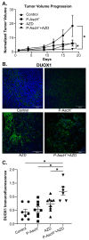
DNMT inhibitors combined with P-AscH decrease tumor volume and increase DUOX1 expression in vivo. Athymic nude mice with heterotopic MIA PaCa-2 xenografts were treated with intraperitoneal normal saline (4 g/kg, 1 M, daily), AZD (1 g/kg, three times weekly), P-AscH (4 g/kg, daily), or a combination of P-AscH and AZD. Mice were treated for 21 days and tumor volume was measured twice weekly. (A) Tumor growth was significantly inhibited in the combination AZD + P-AscH group compared to the control group and compared to either treatment group alone. Tumor volumes (mm3) were normalized to their starting volumes on treatment day 1 to avoid heterogeneity in the starting tumor volumes. Data represent average tumor volume over 18 d (means ± SEM; * p < 0.05). (B) DUOX1 immunofluorescence staining was performed on xenograft tumor samples. Samples were visualized using a Zeiss Confocal Microscope 40× oil objective. Results show increased DUOX1 immunofluorescence in the AZD treatment groups compared to control. Green staining, DUOX1; blue staining, nuclear topoisomerase-3. Representative images are shown. (C) Quantification demonstrating MFI normalized to nuclear content (means ± SEM; n = 7; * p < 0.05).

Similar articles

Cited by

References

    1. Siegel R.L., Miller K.D., Fuchs H.E., Jemal A. Cancer statistics, 2022. CA Cancer J. Clin. 2022;72:7–33. doi: 10.3322/caac.21708. - DOI - PubMed
    1. Conroy T., Hammel P., Hebbar M., Ben Abdelghani M., Wei A.C., Raoul J.L., Chone L., Francois E., Artru P., Biagi J.J., et al. FOLFIRINOX or Gemcitabine as Adjuvant Therapy for Pancreatic Cancer. N. Engl. J. Med. 2018;379:2395–2406. doi: 10.1056/NEJMoa1809775. - DOI - PubMed
    1. Von Hoff D.D., Ervin T., Arena F.P., Chiorean E.G., Infante J., Moore M., Seay T., Tjulandin S.A., Ma W.W., Saleh M.N., et al. Increased survival in pancreatic cancer with nab-paclitaxel plus gemcitabine. N. Engl. J. Med. 2013;369:1691–1703. doi: 10.1056/NEJMoa1304369. - DOI - PMC - PubMed
    1. Goldstein D., El-Maraghi R.H., Hammel P., Heinemann V., Kunzmann V., Sastre J., Scheithauer W., Siena S., Tabernero J., Teixeira L., et al. nab-Paclitaxel plus gemcitabine for metastatic pancreatic cancer: Long-term survival from a phase III trial. J. Natl. Cancer Inst. 2015;107:dju413. doi: 10.1093/jnci/dju413. - DOI - PubMed
    1. Cameron E., Pauling L. Supplemental ascorbate in the supportive treatment of cancer: Reevaluation of prolongation of survival times in terminal human cancer. Proc. Natl. Acad. Sci. USA. 1978;75:4538–4542. doi: 10.1073/pnas.75.9.4538. - DOI - PMC - PubMed

LinkOut - more resources