. 2023 Aug 29;12(9):1688.
doi: 10.3390/antiox12091688.

Exogenous Melatonin Protects against Oxidative Damage to Membrane Lipids Caused by Some Sodium/Iodide Symporter Inhibitors in the Thyroid

Affiliations

Exogenous Melatonin Protects against Oxidative Damage to Membrane Lipids Caused by Some Sodium/Iodide Symporter Inhibitors in the Thyroid

Aleksandra K Gładysz et al. Antioxidants (Basel). .

Abstract

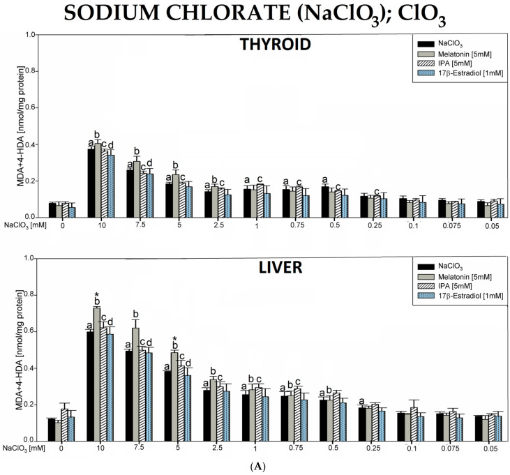
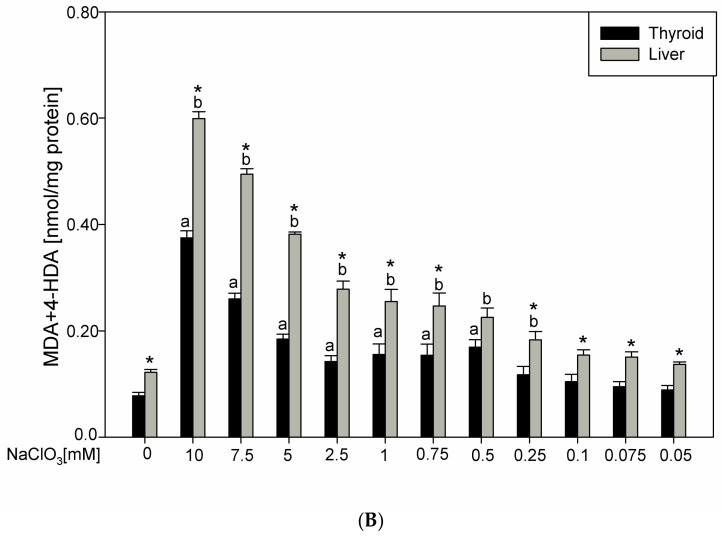
The thyroid gland is the primary site of sodium/iodide symporter (NIS), an intrinsic plasma membrane protein responsible for the active uptake of iodine, which is indispensable for thyroid hormone synthesis. Since exposure of the thyroid to NIS inhibitors can potentially have harmful effects on the entire organism, it is important to investigate the potential protective effects of known antioxidants, such as melatonin and indole-3-propionic acid (IPA), against pro-oxidative action of classic NIS inhibitors. The study aimed to check if and to what extent melatonin and IPA interact with some confirmed NIS inhibitors regarding their effects on oxidative damage to membrane lipids in the thyroid. For comparison with the thyroid gland, in which NIS is typically present, the liver tissue-not possessing NIS-was applied in the present study. Thyroid and liver homogenates were incubated in the presence of tested NIS inhibitors (i.e., NaClO3, NH4SCN, KSeCN, KNO3, NaF, KClO4, and BPA) in different ranges of concentrations with/without melatonin (5 mM) or IPA (5 mM). The malondialdehyde+4-hydroxyalkenals (MDA + 4-HDA) concentration (LPO index) was measured spectrophotometrically. NaClO3 increased LPO in the thyroid and in the liver, but these pro-oxidative effects were not prevented by either melatonin or IPA. Instead, pro-oxidative effects of NH4SCN observed in both tissues were prevented by both indole substances. KSeCN and NaF increased LPO only in the thyroid, and these pro-oxidative effects were prevented by melatonin and IPA. KNO3, KClO4, and BPA did not increase LPO, which can be due to their low concentrations resulting from restricted solubility. In conclusion, as melatonin prevented oxidative damage to membrane lipids in the thyroid caused by some sodium/iodide symporter inhibitors, this indoleamine shoud be considered as a potential protective agent when produced appropriately in living organisms but also as an exogenous substance recommended to individuals overexposed to NIS inhibitors.

Keywords: lipid peroxidation; melatonin; sodium/iodide symporter inhibitors; thyroid.


Conflict of interest statement

The authors declare no conflict of interest.

Figures

Figure 1
Figure 1
(A). Lipid peroxidation (LPO), measured as the level of malondialdehyde + 4-hydroxyalkenals (MDA + 4-HDA) in porcine thyroid or liver homogenates, incubated in the presence of sodium chlorate (NaClO3) (10.0, 7.5, 5.0, 2.5, 1.0, 0.75, 0.5, 0.25, 0.1, 0.075, and 0.05 mM) alone (black bars), or NaClO3 with melatonin (5 mM) (grey bars) or NaClO3 with indole-3-propionic acid (IPA) (5 mM) (striped bars) or NaClO3 with 17β-estradiol (1 mM) (dot bars). LPO level is expressed in nmol/mg protein. Data are presented as mean ± SE (error bars). a p < 0.05 vs. control (without any substance); b p < 0.05 vs. melatonin (5 mM); c p < 0.05 vs. indole-3-propionic acid (5 mM); d p < 0.05 vs. 17β-estradiol (1 mM); * p < 0.05 vs. NaClO3 in the same concentration. (B). Lipid peroxidation (LPO), measured as the level of malondialdehyde + 4-hydroxyalkenals (MDA + 4-HDA), in porcine thyroid (black bars) or liver (grey bars) homogenates. Homogenates were incubated in the presence of sodium chloride (NaClO3) (10.0, 7.5, 5.0, 2.5, 1.0, 0.75, 0.5, 0.25, 0.1, 0.075, and 0.05 mM) used to induce lipid peroxidation. LPO level is expressed in nmol/mg protein. Data are presented as mean ± SE (error bars). a p < 0.05 vs. control in the thyroid; b p < 0.05 vs. control in the liver; * p < 0.05 vs. NaClO3 in the same concentration in the other tissue.
Figure 1
Figure 1
(A). Lipid peroxidation (LPO), measured as the level of malondialdehyde + 4-hydroxyalkenals (MDA + 4-HDA) in porcine thyroid or liver homogenates, incubated in the presence of sodium chlorate (NaClO3) (10.0, 7.5, 5.0, 2.5, 1.0, 0.75, 0.5, 0.25, 0.1, 0.075, and 0.05 mM) alone (black bars), or NaClO3 with melatonin (5 mM) (grey bars) or NaClO3 with indole-3-propionic acid (IPA) (5 mM) (striped bars) or NaClO3 with 17β-estradiol (1 mM) (dot bars). LPO level is expressed in nmol/mg protein. Data are presented as mean ± SE (error bars). a p < 0.05 vs. control (without any substance); b p < 0.05 vs. melatonin (5 mM); c p < 0.05 vs. indole-3-propionic acid (5 mM); d p < 0.05 vs. 17β-estradiol (1 mM); * p < 0.05 vs. NaClO3 in the same concentration. (B). Lipid peroxidation (LPO), measured as the level of malondialdehyde + 4-hydroxyalkenals (MDA + 4-HDA), in porcine thyroid (black bars) or liver (grey bars) homogenates. Homogenates were incubated in the presence of sodium chloride (NaClO3) (10.0, 7.5, 5.0, 2.5, 1.0, 0.75, 0.5, 0.25, 0.1, 0.075, and 0.05 mM) used to induce lipid peroxidation. LPO level is expressed in nmol/mg protein. Data are presented as mean ± SE (error bars). a p < 0.05 vs. control in the thyroid; b p < 0.05 vs. control in the liver; * p < 0.05 vs. NaClO3 in the same concentration in the other tissue.
Figure 2
Figure 2
(A). Lipid peroxidation (LPO), measured as the level of malondialdehyde + 4-hydroxyalkenals (MDA + 4-HDA) in porcine thyroid or liver homogenates, incubated in the presence of ammonium thiocyanate (NH4SCN) (1500, 1250, 1000, 750, 500, 250, 100, 75, 50, 25, and 10 mM) alone (black bars), or NH4SCN plus melatonin (5 mM) (grey bars) or NH4SCN plus indole-3-propionic acid (IPA) (5 mM) (striped bars). LPO level is expressed in nmol/mg protein. Data are presented as mean ± SE (error bars). a p < 0.05 vs. control (without any substance); * p < 0.05 vs. NH4SCN in the same concentration. (B). Lipid peroxidation (LPO), measured as the level of malondialdehyde + 4-hydroxyalkenals (MDA + 4-HDA) in porcine thyroid (black bars) or liver (grey bars) homogenates. Homogenates were incubated in the presence of ammonium thiocyanate (NH4SCN) (1500, 1250, 1000, 750, 500, 250, 100, 75, 50, 25, and 10 mM) used to induce lipid peroxidation. LPO level is expressed in nmol/mg protein. Data are presented as mean ± SE (error bars). a p < 0.05 vs. control in the thyroid; b p < 0.05 vs. control in the liver; * p < 0.05 vs. NH4SCN in the same concentration in the other tissue.
Figure 3
Figure 3
Lipid peroxidation (LPO), measured as the level of malondialdehyde + 4-hydroxyalkenals (MDA + 4-HDA) in porcine thyroid or liver homogenates, incubated in the presence of potassium selenocyanate (KSeCN) (500, 250, 100, 75, 50, 25, 10, 7.5, 5.0, 2.5, and 1.0 mM) alone (black bars), or KSeCN plus melatonin (5 mM) (grey bars) or KSeCN plus indole-3-propionic acid (IPA) (5 mM) (striped bars). LPO level is expressed in nmol/mg protein. Data are presented as mean ± SE (error bars). a p < 0.05 vs. control (without any substance); * p < 0.05 vs. KSeCN in the same concentration.
Figure 4
Figure 4
Lipid peroxidation (LPO), measured as the level of malondialdehyde + 4-hydroxyalkenals (MDA + 4-HDA) in porcine thyroid or liver homogenates, incubated in the presence of potassium nitrate (KNO3) (10.0, 7.5, 5.0, 2.5, 1.0, 0.75, 0.5, 0.25, 0.1 0.075, and 0.05 mM) or—in case of thyroid tissue—KNO3 plus melatonin (5 mM) (grey bars) or KNO3 plus indole-3-propionic acid (IPA) (5 mM) (striped bars). LPO level is expressed in nmol/mg protein. Data are presented as mean ± SE (error bars). No significant differences were found.
Figure 5
Figure 5
Lipid peroxidation (LPO), measured as the level of malondialdehyde + 4-hydroxyalkenals (MDA + 4-HDA) in porcine thyroid or liver homogenates, incubated in the presence of sodium fluoride (NaF) (100, 75, 50, 25, 10, 7.5, 5.0, 2.5, 1.0, 0.75, and 0.5 mM) alone (black bars), or—in case of thyroid tissue—NaF plus melatonin (5 mM) (grey bars) or NaF plus indole-3-propionic acid (IPA) (5 mM) (striped bars). LPO level is expressed in nmol/mg protein. Data are presented as mean ± SE (error bars). a p < 0.05 vs. control (without any substance); * p < 0.05 vs. NaF in the same concentration.
Figure 6
Figure 6
Lipid peroxidation (LPO), measured as the level of malondialdehyde + 4-hydroxyalkenals (MDA + 4-HDA), in porcine thyroid or liver homogenates, incubated in the presence of potassium perchlorate (KClO4) (10.0, 7.5, 5.0, 2.5, 1.0, 0.75, 0.5, 0.25, 0.1, 0.075, and 0.05 mM) or—in case of thyroid tissue—KClO4 plus melatonin (5 mM) (grey bars) or KClO4 plus indole-3-propionic acid (IPA) (5 mM) (striped bars). LPO level is expressed in nmol/mg protein. Data are presented as mean ± SE (error bars).
Figure 7
Figure 7
Lipid peroxidation (LPO), measured as the level of malondialdehyde + 4-hydroxyalkenals (MDA + 4-HDA) in porcine thyroid or liver homogenates, incubated in the presence of bisphenol A (BPA) (2.0, 1.75, 1.5, 1.25, 1.0, 0.75, 0.5, 0.25, 0.1, 0.075, and 0.05 mM) alone (black bars) or—in case of thyroid tissue—BPA plus melatonin (5 mM) (grey bars) or BPA plus indole-3-propionic acid (IPA) (5 mM) (striped bars). LPO level is expressed in nmol/mg protein. Data are presented as mean ± SE (error bars).

Similar articles

Cited by

References

    1. Sorrenti S., Baldini E., Pironi D., Lauro A., D’Orazi V., Tartaglia F., Tripodi D., Lori E., Gagliardi F., Praticò M., et al. Iodine: Its Role in Thyroid Hormone Biosynthesis and Beyond. Nutrients. 2021;13:4469. doi: 10.3390/nu13124469. - DOI - PMC - PubMed
    1. Riesco-Eizaguirre G., Santisteban P., De la Vieja A. The complex regulation of NIS expression and activity in thyroid and extrathyroidal tissues. Endocr.-Relat. Cancer. 2021;28:T141–T165. doi: 10.1530/ERC-21-0217. - DOI - PubMed
    1. Serrano-Nascimento C., Nunes M.T. Perchlorate, nitrate, and thiocyanate: Environmental relevant NIS-inhibitors pollutants and their impact on thyroid function and human health. Front. Endocrinol. 2022;13:995503. doi: 10.3389/fendo.2022.995503. - DOI - PMC - PubMed
    1. Karbownik-Lewińska M., Stępniak J., Iwan P., Lewiński A. Iodine as a potential endocrine disruptor—A role of oxidative stress. Endocrine. 2022;78:219–240. doi: 10.1007/s12020-022-03107-7. - DOI - PMC - PubMed
    1. USEPA . Inorganic Chlorates Facts. US Environmental Protection Agency; Washington, DC, USA: 2008. pp. 1–6. EPA 738-F-08-001.

LinkOut - more resources