. 2023 Sep 15;12(9):1767.
doi: 10.3390/antiox12091767.

Antifungal Action of Arabidopsis thaliana TCP21 via Induction of Oxidative Stress and Apoptosis

Affiliations

Antifungal Action of Arabidopsis thaliana TCP21 via Induction of Oxidative Stress and Apoptosis

Seong-Cheol Park et al. Antioxidants (Basel). .

Abstract

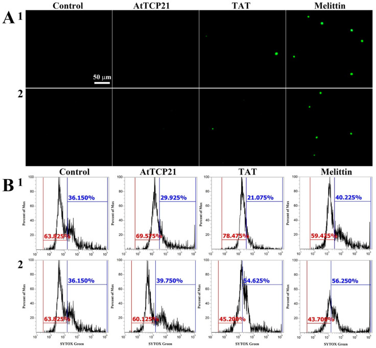
The realm of antimicrobial proteins in plants is extensive but remains relatively uncharted. Understanding the mechanisms underlying the action of plant antifungal proteins (AFPs) holds promise for antifungal strategies. This study aimed to bridge this knowledge gap by comprehensively screening Arabidopsis thaliana species to identify novel AFPs. Using MALDI-TOF analysis, we identified a member of the TEOSINTE BRANCHED1/CYCLOIDEA/PROLIFERATING CELL FACTOR1 (TCP) family of transcription factors as a novel AFP, A. thaliana TCP21 (AtTCP21; accession number NP_196450). Bacterially purified recombinant AtTCP21 inhibited the growth of various pathogenic fungal cells. AtTCP21 was more potent than melittin, a well-known AFP, in combating Colletotrichum gloeosporioides. Growth inhibition assays against various fungal pathogens and yeasts confirmed the pH-dependent antimicrobial activity of AtTCP21. Without inducing any membrane alterations, AtTCP21 penetrates the fungal cell wall and membrane, where it instigates a repressive milieu for fungal cell growth by generating intracellular reactive oxygen species and mitochondrial superoxides; resulting in morphological changes and apoptosis. Our findings demonstrate the redox-regulating effects of AtTCP21 and point to its potential as an antimicrobial agent.

Keywords: TCP; antifungal protein; apoptosis; oxidative stress; reactive oxygen species.


Conflict of interest statement

The authors declare no conflict of interest.

Figures

Figure 1
Figure 1
Characterization of recombinant AtTCP21 based on (A) size distribution, (B) structure, and (C) antifungal activity. (A) Size exclusion chromatography of recombinant AtTCP21 on Superdex S-200 column. Bacterially expressed recombinant AtTCP21 protein was resolved with 12% SDS-PAGE (inset). (B) Purified AtTCP21 was observed under a transmission electron microscope. (C) Inhibitory action of AtTCP21 on the fungal growth. The (1) C. gloeosporioides and (2) F. oxysporum spores were incubated with two-fold diluted concentrations of AtTCP21 at pH 5.5 or 7.2 for 24 h. The treated spores (20 µL) were plated on agar plates and incubated for 48 h.
Figure 2
Figure 2
The effect of AtTCP21 and melittin on cell viability. (A) C. gloeosporioides and (B) F. graminearum cells treated with AtTCP21 or melittin were stained using the live/dead cell assay. FUN-1 dye stains the intracellular intravacuolar structures, and Calcofluor white M2R labels cell-wall chitin, emitting red-orange and blue fluorescence, respectively.
Figure 3
Figure 3
Cellular distribution of Flamma 675-labeled (A) AtTCP21 and (B) melittin in C. gloeosporioides. After incubation of dye-labeled proteins with C. gloeosporioides for 4 h, the washed and fixed fungal cells were observed under confocal laser scanning microscopy (CLSM).
Figure 4
Figure 4
SYTOX Green uptake in (1) C. gloeosporioides and (2) F. graminearum conidial cells. AtTCP21, TAT, and melittin proteins were incubated with fungal conidia for 4 h, followed by the addition of SYTOX Green dye. After further incubation for 15 min, cells were (A) observed under a fluorescence microscope or (B) analyzed via flow cytometry.
Figure 5
Figure 5
Generation of intracellular ROS in response to AtTCP21 in the hyphae and conidia of (a) C. gloeosporioides, (b) F. graminearum, and (c) F. solani cells. Panel (B) shows the quantification of the fluorescence data (A). Statistical significance was analyzed via the one-way ANOVA test, compared to control (** p < 0.01). The bar is 100 μm.
Figure 6
Figure 6
AtTCP21-induced production of mitochondrial superoxide in C. gloeosporioides cells. After incubation of AtTCP21 (IC50 concentration) or H2O2 (1 mM) for 6 h in conidial cells, they were observed under fluorescence microscope (A) and analyzed using flow cytometry (B).
Figure 7
Figure 7
Quantification of cytochrome c release (A), levels of reduced GSH, and oxidized GSSG (B) in AtTCP21- and melittin-treated C. gloeosporioides cells (** p < 0.01).
Figure 8
Figure 8
Induction of apoptosis and caspase 3/7 activation in AtTCP21-treated C. gloeosporioides cells. C. gloeosporioides conidia were incubated with AtTCP21 or melittin at IC50 concentration for 8 h and stained with Annexin V-Flamma 488/PI (A) or caspase-3-7/SYTOX AADvanced dyes (B), flowed via flow cytometry analysis.
Figure 9
Figure 9
Nuclear staining of C. gloeosporioides conidial cells that were (A) untreated, (B) AtTCP21- or (C) melittin-treated. After incubating with AtTCP21 or melittin (IC50 concentration, 6 h), the cells were stained with Hoechst 33342 dye for 20 min and observed under a fluorescence microscope.
Figure 10
Figure 10
Antifungal effects of AtTCP21 and melittin in C. gloeosporioides cells.

Similar articles

Cited by

References

    1. Laxminarayan R., Duse A., Wattal C., Zaidi A.K., Wertheim H.F., Sumpradit N., Vlieghe E., Hara G.L., Gould I.M., Goossens H., et al. Antibiotic resistance-the need for global solutions. Lancet Infect. Dis. 2013;13:1057–1098. doi: 10.1016/S1473-3099(13)70318-9. - DOI - PubMed
    1. Erdem Büyükkiraz M., Kesmen Z. Antimicrobial peptides (AMPs): A promising class of antimicrobial compounds. J. Appl. Microbiol. 2022;132:1573–1596. doi: 10.1111/jam.15314. - DOI - PubMed
    1. Stotz H.U., Thomson J.G., Wang Y. Plant defensins: Defense, development and application. Plant Signal Behav. 2009;4:1010–1012. doi: 10.4161/psb.4.11.9755. - DOI - PMC - PubMed
    1. Sharma P., Kaur J., Sharma G., Kashyap P. Plant derived antimicrobial peptides: Mechanism of target, isolation techniques, sources and pharmaceutical applications. J. Food Biochem. 2022;46:e14348. doi: 10.1111/jfbc.14348. - DOI - PubMed
    1. Bakare O.O., Gokul A., Fadaka A.O., Wu R., Niekerk L.A., Barker A.M., Keyster M., Klein A. Plant Antimicrobial Peptides (PAMPs): Features, Applications, Production, Expression, and Challenges. Molecules. 2022;27:3703. doi: 10.3390/molecules27123703. - DOI - PMC - PubMed

LinkOut - more resources