. 2023 Sep 18;12(9):1775.
doi: 10.3390/antiox12091775.

Pharmacological Ascorbate Elicits Anti-Cancer Activities against Non-Small Cell Lung Cancer through Hydrogen-Peroxide-Induced-DNA-Damage

Affiliations

Pharmacological Ascorbate Elicits Anti-Cancer Activities against Non-Small Cell Lung Cancer through Hydrogen-Peroxide-Induced-DNA-Damage

Kittipong Sanookpan et al. Antioxidants (Basel). .

Abstract

Non-small cell lung cancer (NSCLC) poses a significant global health burden with unsatisfactory survival rates, despite advancements in diagnostic and therapeutic modalities. Novel therapeutic approaches are urgently required to improve patient outcomes. Pharmacological ascorbate (P-AscH-; ascorbate at millimolar concentration in plasma) emerged as a potential candidate for cancer therapy for recent decades. In this present study, we explore the anti-cancer effects of P-AscH- on NSCLC and elucidate its underlying mechanisms. P-AscH- treatment induces formation of cellular oxidative distress; disrupts cellular bioenergetics; and leads to induction of apoptotic cell death and ultimately reduction in clonogenic survival. Remarkably, DNA and DNA damage response machineries are identified as vulnerable targets for P-AscH- in NSCLC therapy. Treatments with P-AscH- increase the formation of DNA damage and replication stress markers while inducing mislocalization of DNA repair machineries. The cytotoxic and genotoxic effects of P-AscH- on NSCLC were reversed by co-treatment with catalase, highlighting the roles of extracellular hydrogen peroxide in anti-cancer activities of P-AscH-. The data from this current research advance our understanding of P-AscH- in cancer treatment and support its potential clinical use as a therapeutic option for NSCLC therapy.

Keywords: DNA damage; adjuvant; anti-cancer; ascorbic acid; non-small cell lung cancer; oxidative distress; pharmacological ascorbate; pro-oxidant; vitamin C.


Conflict of interest statement

The authors declare no potential conflict of interest. Kittipong Sanookpan, M.Sc. currently holds the position as Research Manager of Nabsolute Co., Ltd. It is important to note that the Nabsolute Co., Ltd. was not involved in the study’s design, data collection, data analysis, data interpretation, manuscript preparation or the decision to publish the findings of this research.

Figures

Figure 1
Figure 1
Pharmacological ascorbate possesses anti-cancer activities against NSCLC cells. (A) P-AscH kills NSCLC cells in dose-dependent fashion. Cells were exposed to P-AscH (0–6 mM; 1 h) and cell viability was evaluated at 24 h after treatments using MTT assay. (B) The concentration of P-AscH required to decrease viability of H23, H292, or H460 by 50% (EC50) was further evaluated on the basis of the dose–response relationship. (C,D) P-AscH dose-dependently reduces clonogenicity of NSCLC cells. Cells were treated with P-AscH (0–4 mM; 1 h) and the ability of cancer cells to form a colony was determined following treatments with clonogenic survival assay (n = 3; mean ± SEM; * p < 0.05; *** p < 0.001; **** p < 0.0001 vs. untreated control).
Figure 2
Figure 2
The generation of extracellular H2O2 is an essential factor for anti-cancer properties of P-AscH against NSCLC cells. (A) The intracellular levels of GSH of NSCLC cells were reduced after P-AscH treatments. Cells were exposed to P-AscH (0–4 mM) for 1 h; and the redox status of NSCLC cells was immediately evaluated by a measurement of an intracellular concentration of GSH. (B,C) P-AscH induces generation of oxidative distress in NSCLC cells via the formation of H2O2. To investigate roles of H2O2, NSCLC cells were treated with P-AscH (4 mM) ± extracellular catalase (200 U/mL) for 1 h. The oxidative status of cells was subsequently determined by flow cytometric analysis using DCFH-DA fluorescent probe. (D,E) The formation of extracellular H2O2 is crucial for cytotoxicity of P-AscH against NSCLC cells. Co-treatment with catalase prevented a decrease in clonogenic survival of NSCLC cells due to P-AscH exposure. The treatment protocols were similar to those described in (B,C) (n = 3; mean ± SEM; * p < 0.01, *** p < 0.001; **** p < 0.0001 vs. untreated control; †††† p < 0.0001 vs. P-AscH).
Figure 3
Figure 3
Pharmacological ascorbate exhausts intracellular storage of ATP and NAD+ via generation of H2O2. (A,B) The intracellular pools of ATP and NAD+ of NSCLC cells were rapidly depleted upon treatment with P-AscH. Cells were incubated with P-AscH (0–4 mM) for 1 h, and the immediate impact on intracellular amounts of ATP (A) and NAD+ pool (B) was observed. (C,D) The disruption of cellular bioenergetics by P-AscH is primarily mediated by formation of extracellular H2O2. Catalase prevents the reduction in ATP levels (C) and NAD+ storage (D) in NSCLC cells following P-AscH treatment. The treatment protocols were similar to 2B and 2C (n = 3; mean ± SEM; ** p < 0.01, *** p < 0.001, **** p < 0.0001 vs. untreated control; †††† p < 0.0001 vs. P-AscH).
Figure 3
Figure 3
Pharmacological ascorbate exhausts intracellular storage of ATP and NAD+ via generation of H2O2. (A,B) The intracellular pools of ATP and NAD+ of NSCLC cells were rapidly depleted upon treatment with P-AscH. Cells were incubated with P-AscH (0–4 mM) for 1 h, and the immediate impact on intracellular amounts of ATP (A) and NAD+ pool (B) was observed. (C,D) The disruption of cellular bioenergetics by P-AscH is primarily mediated by formation of extracellular H2O2. Catalase prevents the reduction in ATP levels (C) and NAD+ storage (D) in NSCLC cells following P-AscH treatment. The treatment protocols were similar to 2B and 2C (n = 3; mean ± SEM; ** p < 0.01, *** p < 0.001, **** p < 0.0001 vs. untreated control; †††† p < 0.0001 vs. P-AscH).
Figure 4
Figure 4
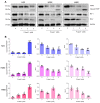
DNA is a susceptible target for treatment of P-AscH. (A) Representative data from Western blot analyses demonstrate that P-AscH treatment leads to a decrease in DNA damage regulators (Chk1 and RPA2); as well as an increase in markers of DNA damage and replication stress following P-AscH exposure (phosphorylation at Ser324 of Chk1, p-Chk1; phosphorylation at Ser33 of RPA2, p-RPA2; phosphorylation at Ser139 of histone H2AX, γ-H2AX). Cells were treated with P-AscH (4 mM; 1 h) and the alterations in DNA damage response system were subsequently determined with Western blot assays at indicated time points (0–24 h after treatments). (B), The relative expression of the indicated proteins compared to the untreated control were calculated from densitometric analysis and normalized to GAPDH expression (n = 3; mean ± SEM; * p < 0.05, ** p < 0.01, *** p < 0.001, **** p < 0.0001 vs. untreated control).
Figure 5
Figure 5
Formation of H2O2 is an essential factor for genotoxicity of P-AscH against NSCLC. (A) Catalase prevents loss of Chk1 and RPA2; and inhibits formation of p-Chk1, p-RPA2, and γ-H2AX. Cells were co-treated with P-AscH (4 mM) and bovine catalase (200 U/mL) for 1 h, then the changes in DNA damage response pathway were determined at 6 h post-treatment with Western blot analyses. Data are representative of three independent studies. (B) The density of each blot was estimated and subsequently normalized to loading control GAPDH. The values were expressed as fold change relative to the untreated control (n = 3; mean ± SEM; * p < 0.05, *** p < 0.001, **** p < 0.0001 vs. untreated control; ††† p < 0.001, †††† p < 0.0001 vs. P-AscH).
Figure 6
Figure 6
Pharmacological ascorbate affects the localization of key components of the DNA damage response pathway in NSCLC cells. (A) Immunofluorescence staining: H460 cells were treated with 4 mM P-AscH, bovine catalase (200 U/mL), or co-treated with P-AscH (4 mM) and catalase (200 U/mL) for 1 h. Then, cells were subjected to immunofluorescence staining with anti-γ-H2AX (green), anti-RAP80 (red), and anti-BRCA1 (yellow) antibodies at 6 h post-treatment to observe protein localization. Cell nuclei were stained with DAPI (blue). Data are representative of three independent studies. Scale bar, 20 µm. (B) The levels of cytoplasmic γ-H2AX per cell were quantified. The data were expressed as fold change relative to the untreated control (n = 3; mean ± SEM; ** p < 0.01 vs. untreated control;  p < 0.05 vs. P-AscH).
Figure 7
Figure 7
Pharmacological ascorbate causes apoptotic cell death in NSCLC via H2O2 production. (A,B) Exposure to P-AscH (1–4 mM; 1 h) induces apoptotic cell death in NSCLC in a dose-dependent fashion. (C,D) The inductions of apoptosis by P-AscH (1–4 mM; 1 h) on NSCLC were prevented by catalase co-treatment. Cells were treated with P-AscH (0–4 mM) ± catalase (200 U/mL) for 1 h. At 24 h after treatments, the modes of cell death were characterized with flow cytometric assay using annexin V/PI co-staining (n = 3; mean ± SEM; * p < 0.05, ** p < 0.01; *** p < 0.001, **** p < 0.0001 vs. untreated control).
Figure 7
Figure 7
Pharmacological ascorbate causes apoptotic cell death in NSCLC via H2O2 production. (A,B) Exposure to P-AscH (1–4 mM; 1 h) induces apoptotic cell death in NSCLC in a dose-dependent fashion. (C,D) The inductions of apoptosis by P-AscH (1–4 mM; 1 h) on NSCLC were prevented by catalase co-treatment. Cells were treated with P-AscH (0–4 mM) ± catalase (200 U/mL) for 1 h. At 24 h after treatments, the modes of cell death were characterized with flow cytometric assay using annexin V/PI co-staining (n = 3; mean ± SEM; * p < 0.05, ** p < 0.01; *** p < 0.001, **** p < 0.0001 vs. untreated control).
Figure 8
Figure 8
Pharmacological ascorbate induces activation of apoptotic signaling cascade on NSCLC. (A) The hallmarks of apoptotic cell death (increased PARP cleavage and reduced expression of anti-apoptotic proteins (Mcl-1 and Bcl-2)) were observed in NSCLC with Western blot analysis at 24 h after treatments with P-AscH (0–4 mM; 1 h). (B) The density of each protein band was measured and normalized to GAPDH. The data were presented as fold change relative to the untreated control (n = 3; mean ± SEM; * p < 0.05, ** p < 0.01; *** p < 0.001, **** p < 0.0001 vs. untreated control).
Figure 9
Figure 9
Pharmacological ascorbate synergistically enhances anti-cancer activities of chemotherapeutic agents against NSCLC. (A) The addition of P-AscH augments the cytotoxicity of gemcitabine (GEM) in H23 cells. (B) The graph illustrates the combination indices (CI) plotted against the fraction affected (Fa), indicating the synergistic interaction between P-AscH and GEM. (C) P-AscH amplifies the anti-cancer activities of docetaxel (DTX) in H292 cells. (D) The CI values between P-AscH/DTX are consistently below 1, suggesting the synergistic effects of this combination. Cells were exposed to varying amounts of chemotherapeutic drugs (either GEM or DTX; 24 h); or P-AscH (1 h); or a combined treatment of respective chemotherapeutic drugs (24 h) followed by P-AscH (1 h). At 24 h post-treatments, cell viability was assessed with MTT assay and combination indices were calculated to assess interaction effect. For combination treatments, the consistent ratio of GEM:P-P-AscH (1:1.5) and DTX: P-AscH (1:8.9) were maintained, respectively. The presented data were derived from three independent experiments.

Similar articles

Cited by

References

    1. Sung H., Ferlay J., Siegel R.L., Laversanne M., Soerjomataram I., Jemal A., Bray F. Global Cancer Statistics 2020: GLOBOCAN Estimates of Incidence and Mortality Worldwide for 36 Cancers in 185 Countries. CA Cancer J. Clin. 2021;71:209–249. doi: 10.3322/caac.21660. - DOI - PubMed
    1. Molina J.R., Yang P., Cassivi S.D., Schild S.E., Adjei A.A. Non-small cell lung cancer: Epidemiology, risk factors, treatment, and survivorship. Mayo Clin. Proc. 2008;83:584–594. doi: 10.1016/S0025-6196(11)60735-0. - DOI - PMC - PubMed
    1. Garon E.B., Hellmann M.D., Rizvi N.A., Carcereny E., Leighl N.B., Ahn M.J., Eder J.P., Balmanoukian A.S., Aggarwal C., Horn L., et al. Five-year overall survival for patients with advanced non-small-cell lung cancer treated with pembrolizumab: Results from the phase I KEYNOTE-001 Study. J. Clin. Oncol. 2019;37:2518–2527. doi: 10.1200/JCO.19.00934. - DOI - PMC - PubMed
    1. Du J., Cullen J.J., Buettner G.R. Ascorbic acid: Chemistry, biology and the treatment of cancer. Biochim. Biophys. Acta. 2012;1826:443–457. doi: 10.1016/j.bbcan.2012.06.003. - DOI - PMC - PubMed
    1. Buranasudja V., Doskey C.M., Gibson A.R., Wagner B.A., Du J., Gordon D.J., Koppenhafer S.L., Cullen J.J., Buettner G.R. Pharmacologic ascorbate primes pancreatic cancer cells for death by rewiring cellular energetics and inducing DNA damage. Mol. Cancer Res. 2019;17:2102–2114. doi: 10.1158/1541-7786.MCR-19-0381. - DOI - PMC - PubMed

LinkOut - more resources