. 2023 Aug 24;10(9):999.
doi: 10.3390/bioengineering10090999.

Enhanced Expansion of Human Pluripotent Stem Cells and Somatic Cell Reprogramming Using Defined and Xeno-Free Culture Conditions

Affiliations

Enhanced Expansion of Human Pluripotent Stem Cells and Somatic Cell Reprogramming Using Defined and Xeno-Free Culture Conditions

Suraj Timilsina et al. Bioengineering (Basel). .

Abstract

Human embryonic stem cells and induced pluripotent stem cells (hPSC) have an unprecedented opportunity to revolutionize the fields of developmental biology as well as tissue engineering and regenerative medicine. However, their applications have been significantly limited by the lack of chemically defined and xeno-free culture conditions. The demand for the high-quality and scaled-up production of cells for use in both research and clinical studies underscores the need to develop tools that will simplify the in vitro culture process while reducing the variables. Here, we describe a systematic study to identify the optimal conditions for the initial cell attachment of hPSC to tissue culture dishes grafted with polymers of N-(3-Sulfopropyl)-N-Methacryloxyethyl-N, N-Dimethylammoniun Betaine (PMEDSAH) in combination with chemically defined and xeno-free culture media. After testing multiple supplements and chemicals, we identified that pre-conditioning of PMEDSAH grafted plates with 10% human serum (HS) supported the initial cell attachment, which allowed for the long-term culture and maintenance of hPSC compared to cells cultured on Matrigel-coated plates. Using this culture condition, a 2.1-fold increase in the expansion of hPSC was observed without chromosomal abnormalities. Furthermore, this culture condition supported a higher reprogramming efficiency (0.37% vs. 0.22%; p < 0.0068) of somatic cells into induced pluripotent stem cells compared to the non-defined culture conditions. This defined and xeno-free hPSC culture condition may be used in obtaining the large populations of hPSC and patient-derived iPSC required for many applications in regenerative and translational medicine.

Keywords: PMEDSAH; human embryonic stem cells; human pluripotent stem cells; reprogramming; self-renewal; synthetic polymer grafting.


Conflict of interest statement

LVD declares a conflict of interest.

Figures

Figure 1
Figure 1
Schematic representation of the experimental protocol. PMEDSAH grafted (PMEDSAH-g) TCP dishes were synthesized using a surface-initiated graft polymerization procedure. The dishes were then pre-conditioned/treated with 10% human serum (HSt) (v/v DMEM/F12) and separated into two sets of 10% HSt-PMEDSAH-g dishes. The first set (W10% HSt-PMEDSAH-g) was used right after its preparation for culturing hPSC for 20 consecutive passages (wet-W). The other set (D10% HSt-PMEDSAH-g) was air dried and wrapped with parafilm, stored at 4 °C until their use for cell culture (dry-D), and used for the weekly passaging of hPSC for 13 consecutive passages. In both conditions, the cells were characterized for self-renewal and pluripotency.
Figure 2
Figure 2
Culture and expansion of hPSC on W10% HSt-PMEDSAH-g dishes. Fresh PMEDSAH-g dishes treated with 10% HS (W10% HSt-PMEDSAH-g) in DMEM/F12 gave better results for promoting and stabilizing cell attachment and long-term culture of undifferentiated hPSC compared to cells cultured on Matrigel-coated dishes. (A) Representative micrographs of hPSC colonies cultured on Matrigel-coated plates and W10% HSt-PMEDSAH-g. On the right side, respective alkaline phosphatase (AP) stained colonies. (B) A plot of undifferentiated colony number (ratio) and total cell number (ratio) compared to the control group (Matrigel-coated dishes) indicating that W10% HSt-PMEDSAH-g lead to a higher number of undifferentiated colonies and the total number of cells on week 1. (C) Representative micrographs of hPSC colonies cultured on W10% HSt-PMEDSAH-g and Matrigel-coated plates during week 0 and week 5. The graph to the right indicates the theoretical yield of the total number of cells counted and compared from colonies cultured on W10% HSt-PMEDSAH-g and Matrigel-coated dishes during weekly passaging until passage 5. Scale bars, 1000 µm. * p < 0.05, (n = 3; unpaired t test). Error bars in graphs represent the SEM of the group.
Figure 3
Figure 3
Characterization of hPSC after long-term culture on W10% HSt-PMEDSAH-g dishes. hPSC cultured on W10% HSt-PMEDSAH-g dishes for 20 consecutive passages, maintaining self-renewal and pluripotency. (A) Representative micrographs of immunocytochemistry every 5 weeks showing strong positive staining for human pluripotency markers NANOG, OCT4, SOX2, TRA1-60, and TRA-181. (B) Dot plots of hPSC after 20 passages of culture on W10% HSt-PMEDSAH-g dishes, showing more than 99% co-expression between SSEA3/OCT4 and SSEA4/OCT4. Isotype controls for the respective antibodies were used. The threshold/gate was set to a maximum of 0.8% positive cells in the unstained control and every signal above was counted as a positive signal. FMO controls were used to assess the spread of the fluorochrome into the missing channel and the gates were set accordingly. (C) Pluripotency was confirmed by qPCR hPSC ScoreCard assay quantifying the self-renewal and trilineage differentiation potential of passage 20 hPSC cultured on W10% HSt-PMEDSAH-g and 10 days old EB developed from the same pool of cells. (D) G-banding karyogram of cells cultured on W10% HSt-PMEDSAH-g dishes at passage 20 confirming genetic integrity. Scale bars, 200 µm.
Figure 4
Figure 4
Reprogramming of human gingival fibroblasts into induced PSC (iPSC) on W10% HSt-PMEDSAH-g dishes. Human gingival fibroblasts were reprogrammed into iPSC on Matrigel-coated dishes and on W10% HSt-PMEDSAH-g dishes. (A) Representative transmitted light micrographs showing fibroblast morphology before and during the development of hiPSC colonies, Scale bars, 1000 µm. (B) Representative micrographs of hiPSC developed on W10% HSt-PMEDSAH-g plates after immunocytochemistry staining with pluripotency-associated markers. DAPI was used as nuclear marker. Scale bars, 200 µm. (C) Representative histograms of hiPSC developed on W10% HSt-PMEDSAH-g dishes showing the expression of cell surface pluripotency-associated markers. Isotype controls for the respective antibodies were used. The threshold/gate was set to a maximum of 0.8% positive cells in the unstained control and every signal above was counted as a positive signal. (D) qPCR analysis for the expression of markers corresponding to all germ lineages showing trilineage differentiation potentiality of 10 days old EBs obtained from hiPSC developed on W10% HSt-PMEDSAH-g dishes and compared to undifferentiated hPSC. * p < 0.05, ** p < 0.005, *** p < 0.0005 (n = 3; unpaired t test). Error bars in graphs represent the SEM of the group.
Figure 5
Figure 5
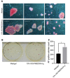
Improved reprogramming efficiency of human gingival fibroblasts into hiPSC on freshly prepared 10% HSt-PMEDSAH-g dishes.Human gingival fibroblasts were reprogrammed into iPSC on W10% HSt-PMEDSAH-g plates and Matrigel-coated plates. (A) Representative bright field images of iPSC colonies derived on W10% HSt-PMEDSAH-g and Matrigel-coated dishes after staining for alkaline phosphatase (AP). Scale bars, 5 mm. (B) Representative images of entire colonies in each condition and (C) graph indicating the average (N = 3) AP-positive colonies per culture dish in each condition. * p < 0.05, (n = 3; unpaired t test). Error bars in graphs represent the SEM of the group.

Similar articles

References

    1. Takahashi K., Tanabe K., Ohnuki M., Narita M., Ichisaka T., Tomoda K., Yamanaka S. Induction of pluripotent stem cells from adult human fibroblasts by defined factors. Cell. 2007;131:861–872. doi: 10.1016/j.cell.2007.11.019. - DOI - PubMed
    1. Thomson J.A., Itskovitz-Eldor J., Shapiro S.S., Waknitz M.A., Swiergiel J.J., Marshall V.S., Jones J.M. Embryonic stem cell lines derived from human blastocysts. Science. 1998;282:1145–1147. doi: 10.1126/science.282.5391.1145. - DOI - PubMed
    1. Yu J., Vodyanik M.A., Smuga-Otto K., Antosiewicz-Bourget J., Frane J.L., Tian S., Nie J., Jonsdottir G.A., Ruotti V., Stewart R., et al. Induced pluripotent stem cell lines derived from human somatic cells. Science. 2007;318:1917–1920. doi: 10.1126/science.1151526. - DOI - PubMed
    1. López S.P., Hernández J.O. Advances in stem cell therapy. Adv. Exp. Med. Biol. 2012;741:290–313. - PubMed
    1. Tamarat R., Lataillade J.J., Bey E., Gourmelon P., Benderitter M. Stem cell therapy: From bench to bedside. Radiat. Prot. Dosim. 2012;151:633–639. doi: 10.1093/rpd/ncs160. - DOI - PubMed

LinkOut - more resources