. 2023 Aug 24;10(9):1004.
doi: 10.3390/bioengineering10091004.

Comparison, Analysis, and Molecular Dynamics Simulations of Structures of a Viral Protein Modeled Using Various Computational Tools

Affiliations

Comparison, Analysis, and Molecular Dynamics Simulations of Structures of a Viral Protein Modeled Using Various Computational Tools

Hemalatha Mani et al. Bioengineering (Basel). .

Abstract

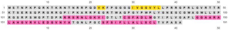
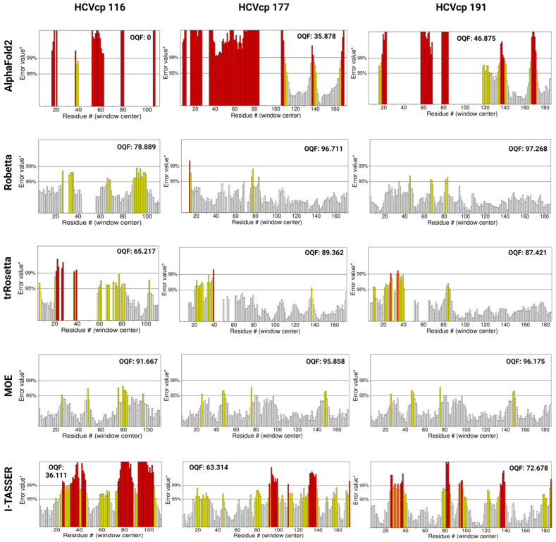
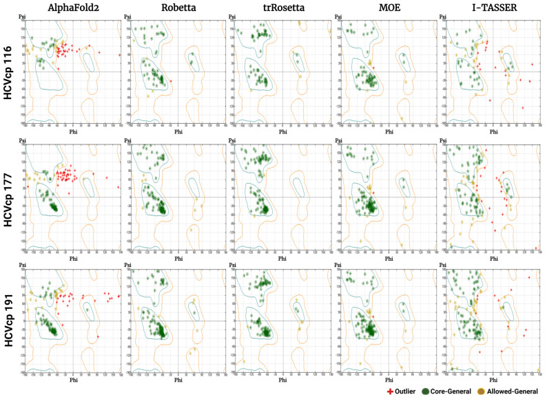
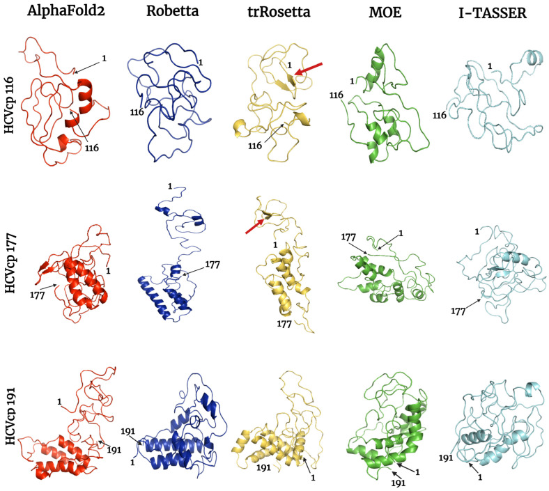
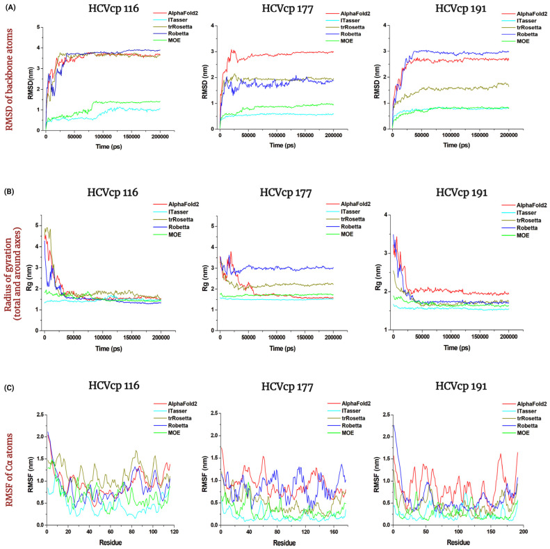
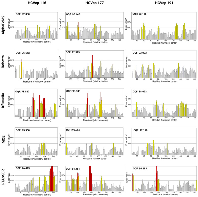
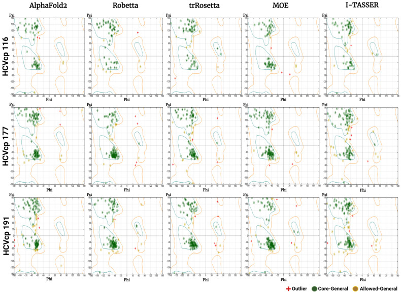
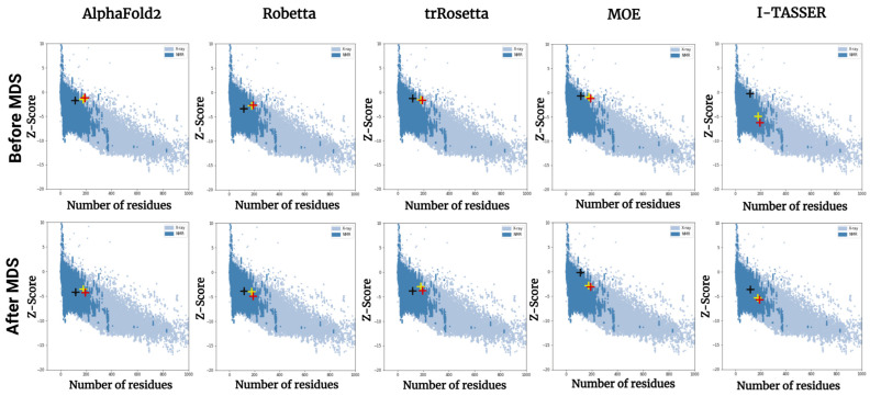
The structural analysis of proteins is a major domain of biomedical research. Such analysis requires resolved three-dimensional structures of proteins. Advancements in computer technology have led to progress in biomedical research. In silico prediction and modeling approaches have facilitated the construction of protein structures, with or without structural templates. In this study, we used three neural network-based de novo modeling approaches-AlphaFold2 (AF2), Robetta-RoseTTAFold (Robetta), and transform-restrained Rosetta (trRosetta)-and two template-based tools-the Molecular Operating Environment (MOE) and iterative threading assembly refinement (I-TASSER)-to construct the structure of a viral capsid protein, hepatitis C virus core protein (HCVcp), whose structure have not been fully resolved by laboratory techniques. Templates with sufficient sequence identity for the homology modeling of complete HCVcp are currently unavailable. Therefore, we performed domain-based homology modeling for MOE simulations. The templates for each domain were obtained through sequence-based searches on NCBI and the Protein Data Bank. Then, the modeled domains were assembled to construct the complete structure of HCVcp. The full-length structure and two truncated forms modeled using various computational tools were compared. Molecular dynamics (MD) simulations were performed to refine the structures. The root mean square deviation of backbone atoms, root mean square fluctuation of Cα atoms, and radius of gyration were calculated to monitor structural changes and convergence in the simulations. The model quality was evaluated through ERRAT and phi-psi plot analysis. In terms of the initial prediction for protein modeling, Robetta and trRosetta outperformed AF2. Regarding template-based tools, MOE outperformed I-TASSER. MD simulations resulted in compactly folded protein structures, which were of good quality and theoretically accurate. Thus, the predicted structures of certain proteins must be refined to obtain reliable structural models. MD simulation is a promising tool for this purpose.

Keywords: AF2; HCV core protein; I-TASSER; MD simulation; MOE; Robetta-RoseTTAFold; homology modeling; trRosetta.


Conflict of interest statement

The authors declare no conflict of interest.

Figures

Figure 1
Figure 1
Tertiary structures of varying lengths of HCVcp (HCVcp 116, HCVcp 177, and HCVcp 191) predicted using AF2 (red), Robetta (blue), trRosetta (yellow), MOE (green), and I-TASSER (cyan). The positions of the first and last amino acids from the N-terminal of the protein are indicated. The red arrows indicate β-strands, which are also observed in the predicted secondary structures.
Figure 2
Figure 2
The AA sequence and secondary structure prediction of HCVcp 191 obtained by using PSIPRED 4.0. Numbers in the figure indicate the positions of amino acids from the N-terminal of the protein. Strands, helices, and coils are indicated with yellow, pink, and grey, respectively.
Figure 3
Figure 3
ERRAT plots for HCVcp structures with varying lengths (HCVcp 116, HCVcp 177, and HCVcp 191). The models were predicted using AF2, Robetta, trRosetta, MOE, and I-TASSER. The plots show two lines (on the error axis), which indicate the confidence level at which regions exceeding the error value can be rejected. The regions that can be rejected at the 95% confidence level are in yellow and those that can be rejected at the 99% confidence level (99% confidence in being erroneous) are in red. OQF, overall quality factor.
Figure 4
Figure 4
Phi–psi plots for the HCVcp 116, HCVcp 177, and HCVcp 191 models constructed using AF2, Robetta, trRosetta, MOE, and I-TASSER. The cyan outline corresponds to most favored regions (conformations with no steric hindrance). The orange outline corresponds to allowed regions. The regions outside the orange outline are disallowed regions (sterically forbidden regions).
Figure 5
Figure 5
Final frames of the refined HCVcp 116, HCVcp 177, and HCVcp 191 models. The models were constructed using AF2 (red), Robetta (blue), trRosetta (yellow), MOE (green), and I-TASSER (cyan) and subsequently subjected to MD simulations. The positions of the first and last amino acids from the N-terminal of the protein are indicated. The red arrows indicate β-strands, which were also observed in the predicted secondary structures.
Figure 6
Figure 6
MD simulation trajectories of (A) RMSD of backbone atoms, (B) radius of gyration, and (C) RMSF of Cα atoms for the models of HCVcp 116, HCVcp 177, and HCVcp 191 during the 200 ns MD simulations. The red, blue, dark yellow, green, and cyan lines indicate the values for the models constructed using AF2, Robetta, trRosetta, MOE, and I-TASSER, respectively.
Figure 7
Figure 7
ERRAT plots for the refined HCVcp 116, HCVcp 177, and HCVcp 191 models. The models were predicted using AF2, Robetta, trRosetta, MOE, and I-TASSER and subsequently subjected to 200 ns MD simulations. The plots show two lines (on the error axis), which indicate the confidence level at which regions exceeding the error value can be rejected. The regions that can be rejected at the 95% confidence level are in yellow, whereas those that can be rejected at the 99% confidence level (99% confidence in being erroneous) are in red. OQF, overall quality factor.
Figure 8
Figure 8
Phi–psi plots of the refined HCVcp 116, HCVcp 177, and HCVcp 191 models. The models were constructed using AF2, Robetta, trRosetta, MOE, and I-TASSER and subsequently subjected to 200 ns MD simulations. The cyan outline corresponds to the most favored regions (conformations with no steric hindrance). The orange outline corresponds to the allowed regions. The regions outside the orange outline are disallowed regions (sterically forbidden regions).
Figure 9
Figure 9
Results of the ProSA-web-based validation of all HCVcp models before (upper panels) and after (lower panels) MD simulations. The ProSA-web z-scores of all available PDB protein structures determined through X-ray crystallography (light blue) or NMR spectroscopy (dark blue) with respect to length. The black, yellow, and red crosses indicate the z-scores of the HCVcp 116, HCVcp 177, and HCVcp 191 models, respectively. ProSA, Protein Structure Analysis.

Similar articles

Cited by

References

    1. Ornes S. Researchers turn to deep learning to decode protein structures. Proc. Natl. Acad. Sci. USA. 2022;119:e2202107119. doi: 10.1073/pnas.2202107119. - DOI - PMC - PubMed
    1. Pakhrin S.C., Shrestha B., Adhikari B., Kc D.B. Deep learning-based advances in protein structure prediction. Int. J. Mol. Sci. 2021;22:5553. doi: 10.3390/ijms22115553. - DOI - PMC - PubMed
    1. Pearce R., Zhang Y. Deep learning techniques have significantly impacted protein structure prediction and protein design. Curr. Opin. Struct. Biol. 2021;68:194–207. doi: 10.1016/j.sbi.2021.01.007. - DOI - PMC - PubMed
    1. Pearce R., Zhang Y. Toward the solution of the protein structure prediction problem. J. Biol. Chem. 2021;297:100870. doi: 10.1016/j.jbc.2021.100870. - DOI - PMC - PubMed
    1. Cramer P. AlphaFold2 and the future of structural biology. Nat. Struct. Mol. Biol. 2021;28:704–705. doi: 10.1038/s41594-021-00650-1. - DOI - PubMed

LinkOut - more resources