

Recent Advancements in Glaucoma Surgery-A Review
- PMID: 37760198
- PMCID: PMC10525614
- DOI: 10.3390/bioengineering10091096
Recent Advancements in Glaucoma Surgery-A Review
Abstract
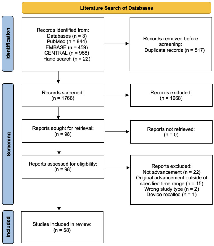
Surgery has long been an important treatment for limiting optic nerve damage and minimising visual loss in patients with glaucoma. Numerous improvements, modifications, and innovations in glaucoma surgery over recent decades have improved surgical safety, and have led to earlier and more frequent surgical intervention in glaucoma patients at risk of vision loss. This review summarises the latest advancements in trabeculectomy surgery, glaucoma drainage device (GDD) implantation, and minimally invasive glaucoma surgery (MIGS). A comprehensive search of MEDLINE, EMBASE, and CENTRAL databases, alongside subsequent hand searches-limited to the past 10 years for trabeculectomy and GDDs, and the past 5 years for MIGS-yielded 2283 results, 58 of which were included in the final review (8 trabeculectomy, 27 GDD, and 23 MIGS). Advancements in trabeculectomy are described in terms of adjunctive incisions, Tenon's layer management, and novel suturing techniques. Advancements in GDD implantation pertain to modifications of surgical techniques and devices, novel methods to deal with postoperative complications and surgical failure, and the invention of new GDDs. Finally, the popularity of MIGS has recently promoted modifications to current surgical techniques and the development of novel MIGS devices.
Keywords: device; eye; glaucoma; glaucoma tube shunts; minimally invasive glaucoma surgery; trabeculectomy.
Conflict of interest statement
B.C.H.A has received speaker’s honoraria from Alcon, Inc., research support and speaker’s honoraria from Glaukos Corporation, and speaker’s honoraria from Santen Pharmaceutical Asia Pte. Ltd. S.D. has received travel support from New World Medical.
Figures
Similar articles
-
Trends and Usage Patterns of Minimally Invasive Glaucoma Surgery in the United States: IRIS® Registry Analysis 2013-2018.Ophthalmol Glaucoma. 2021 Nov-Dec;4(6):558-568. doi: 10.1016/j.ogla.2021.03.012. Epub 2021 Apr 6. Ophthalmol Glaucoma. 2021. PMID: 33831643
-
[MINIMALLY INVASIVE GLAUCOMA SURGERIES].Harefuah. 2019 Jan;158(1):60-64. Harefuah. 2019. PMID: 30663296 Review. Hebrew.
-
A Comprehensive Review of Recent Advances in Minimally Invasive Glaucoma Surgery: Current Trends and Future Directions.Cureus. 2024 Jul 24;16(7):e65236. doi: 10.7759/cureus.65236. eCollection 2024 Jul. Cureus. 2024. PMID: 39184647 Free PMC article. Review.
-
Comparison of mitomycin C trabeculectomy, glaucoma drainage device implantation, and laser neodymium:YAG cyclophotocoagulation in the management of intractable glaucoma after penetrating keratoplasty.Ophthalmology. 1998 Aug;105(8):1550-6. doi: 10.1016/S0161-6420(98)98046-0. Ophthalmology. 1998. PMID: 9709773
-
Differences in the Surgical Outcomes of Glaucoma Surgery in Patients of African Caribbean Descent.Curr Eye Res. 2022 Dec;47(12):1567-1577. doi: 10.1080/02713683.2022.2126859. Epub 2022 Oct 19. Curr Eye Res. 2022. PMID: 36214781
Cited by
-
Challenging glaucoma with emerging therapies: an overview of advancements against the silent thief of sight.Front Med (Lausanne). 2025 Mar 26;12:1527319. doi: 10.3389/fmed.2025.1527319. eCollection 2025. Front Med (Lausanne). 2025. PMID: 40206485 Free PMC article. Review.
-
Fighting Bleb Fibrosis After Glaucoma Surgery: Updated Focus on Key Players and Novel Targets for Therapy.Int J Mol Sci. 2025 Mar 5;26(5):2327. doi: 10.3390/ijms26052327. Int J Mol Sci. 2025. PMID: 40076946 Free PMC article.
-
Three techniques for 360-degree gonioscopy-assisted transluminal trabeculotomy with iTrack advance.Am J Ophthalmol Case Rep. 2024 Oct 5;36:102192. doi: 10.1016/j.ajoc.2024.102192. eCollection 2024 Dec. Am J Ophthalmol Case Rep. 2024. PMID: 39435155 Free PMC article.
-
Structural Updates to the Implant and Refill Needle of the Port Delivery Platform.Transl Vis Sci Technol. 2025 Apr 1;14(4):8. doi: 10.1167/tvst.14.4.8. Transl Vis Sci Technol. 2025. PMID: 40192619 Free PMC article.
-
Evaluating the Safety and Efficacy of a Novel Glaucoma Drainage Device in High-Risk Adult Glaucoma Patients: A One-Year Pilot Study.J Clin Med. 2024 Aug 23;13(17):4996. doi: 10.3390/jcm13174996. J Clin Med. 2024. PMID: 39274211 Free PMC article.
References
Publication types
LinkOut - more resources
Full Text Sources
Miscellaneous
