

Morphea, Eosinophilic Fasciitis and Cancer: A Scoping Review
- PMID: 37760419
- PMCID: PMC10526289
- DOI: 10.3390/cancers15184450
Morphea, Eosinophilic Fasciitis and Cancer: A Scoping Review
Abstract
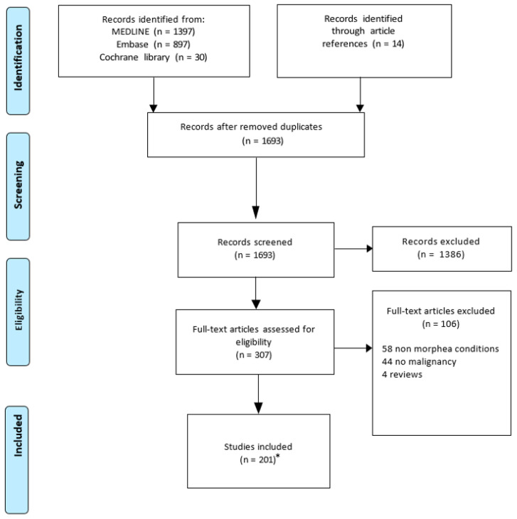
Morphea is an autoimmune fibrotic skin disease. Eosinophilic fasciitis (EF) is considered to belong to the severe spectrum of morphea. We conducted a scoping review assessing the risk of secondary cancer among morphea/EF patients, paraneoplastic morphea/EF and morphea/EF developing secondary to cancer therapy. The search was conducted using MEDLINE, Embase, Cochrane databases for articles published from inception to September 2022 following the Preferred Reporting Items for Systematic reviews and Meta-Analyses for Scoping Reviews (PRISMA-ScR) guidelines with no language or date restrictions. Two hundred and one studies were included. Of these, 32 studies reported on secondary cancer in morphea/EF patients, 45 on paraneoplastic morphea/EF and 125 on cancer-treatment-induced morphea/EF. While the current evidence remains limited, data suggest an increased risk of secondary cutaneous and possibly pancreatic malignancy in morphea patients, particularly the generalized subtype. There were insufficient data for EF. On the other hand, paraneoplastic morphea was anecdotal, whereas several observational studies suggested that ~10% of EF cases may be paraneoplastic, primarily in the context of hematologic malignancies. Radiotherapy-induced morphea is rare, seen in ~0.2% of treated patients and is usually localized to the treatment site, except in patients with pre-existing autoimmunity. While chemotherapy-induced cases are reported, immunotherapy morphea/EF cases are emerging and are preferentially seen with PD-1 and not CTLA-4 inhibitors. This study is limited by the type of articles included (case reports, case series and observational studies), and hence, additional research on this important topic is needed.
Keywords: cancer; chemotherapy; eosinophilic fasciitis; immune checkpoint inhibitors; immunotherapy; localized scleroderma; malignancy; morphea; paraneoplastic; radiation induced scleroderma.
Conflict of interest statement
The authors declare no conflict of interest.
Figures
References
-
- Lagacé F., D’Aguanno K., Prosty C., Laverde-Saad A., Cattelan L., Ouchene L., Oliel S., Genest G., Doiron P., Richer V., et al. The Role of Sex and Gender in Dermatology—From Pathogenesis to Clinical Implications. J. Cutan. Med. Surg. 2023:12034754231177582. doi: 10.1177/12034754231177582. - DOI - PMC - PubMed
-
- Grabell D., Hsieh C., Andrew R., Martires K., Kim A., Vasquez R., Jacobe H. The Role of Skin Trauma in the Distribution of Morphea Lesions: A Cross-Sectional Survey of the Morphea in Adults and Children Cohort IV. J. Am. Acad. Dermatol. 2014;71:493–498. doi: 10.1016/j.jaad.2014.04.009. - DOI - PMC - PubMed
-
- Jacobe H., Ahn C., Arnett F.C., Reveille J.D. Major Histocompatibility Complex Class I and Class II Alleles May Confer Susceptibility to or Protection against Morphea: Findings from the Morphea in Adults and Children Cohort. Arthritis Rheumatol. 2014;66:3170–3177. doi: 10.1002/art.38814. - DOI - PMC - PubMed
Publication types
LinkOut - more resources
Full Text Sources
