Small Renal Masses: Developing a Robust Radiomic Signature
- PMID: 37760532
- PMCID: PMC10527518
- DOI: 10.3390/cancers15184565
Small Renal Masses: Developing a Robust Radiomic Signature
Abstract
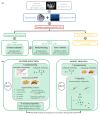
(1) Background and (2) Methods: In this retrospective, observational, monocentric study, we selected a cohort of eighty-five patients (age range 38-87 years old, 51 men), enrolled between January 2014 and December 2020, with a newly diagnosed renal mass smaller than 4 cm (SRM) that later underwent nephrectomy surgery (partial or total) or tumorectomy with an associated histopatological study of the lesion. The radiomic features (RFs) of eighty-five SRMs were extracted from abdominal CTs bought in the portal venous phase using three different CT scanners. Lesions were manually segmented by an abdominal radiologist. Image analysis was performed with the Pyradiomic library of 3D-Slicer. A total of 108 RFs were included for each volume. A machine learning model based on radiomic features was developed to distinguish between benign and malignant small renal masses. The pipeline included redundant RFs elimination, RFs standardization, dataset balancing, exclusion of non-reproducible RFs, feature selection (FS), model training, model tuning and validation of unseen data. (3) Results: The study population was composed of fifty-one RCCs and thirty-four benign lesions (twenty-five oncocytomas, seven lipid-poor angiomyolipomas and two renal leiomyomas). The final radiomic signature included 10 RFs. The average performance of the model on unseen data was 0.79 ± 0.12 for ROC-AUC, 0.73 ± 0.12 for accuracy, 0.78 ± 0.19 for sensitivity and 0.63 ± 0.15 for specificity. (4) Conclusions: Using a robust pipeline, we found that the developed RFs signature is capable of distinguishing RCCs from benign renal tumors.
Keywords: benign; characterization; kidney cancer; malignant; oncocytoma; radiomics; renal cell carcinoma; small renal masses.
Conflict of interest statement
The authors declare no conflict of interest.
Figures


References
-
- «LINEE GUIDA TUMORI DEL RENE». AIOM, 31 December 2021. [(accessed on 29 May 2023)]. Available online: https://www.aiom.it/linee-guida-aiom-2021-tumori-del-rene/
-
- Ljungberg B., Albiges L., Abu-Ghanem Y., Bedke J., Capitanio U., Dabestani S., Fernández-Pello S., Giles R.H., Hofmann F., Hora M., et al. European Association of Urology Guidelines on Renal Cell Carcinoma: The 2022 Update. Eur. Urol. 2022;82:399–410. doi: 10.1016/j.eururo.2022.03.006. - DOI - PubMed
LinkOut - more resources
Full Text Sources
Research Materials

