Review
. 2023 Sep 19;15(18):4629.
doi: 10.3390/cancers15184629.

Changes in the Acetylcholinesterase Enzymatic Activity in Tumor Development and Progression

Affiliations
Review

Changes in the Acetylcholinesterase Enzymatic Activity in Tumor Development and Progression

Benjamín Pérez-Aguilar et al. Cancers (Basel). .

Abstract

Acetylcholinesterase is a well-known protein because of the relevance of its enzymatic activity in the hydrolysis of acetylcholine in nerve transmission. In addition to the catalytic action, it exerts non-catalytic functions; one is associated with apoptosis, in which acetylcholinesterase could significantly impact the survival and aggressiveness observed in cancer. The participation of AChE as part of the apoptosome could explain the role in tumors, since a lower AChE content would increase cell survival due to poor apoptosome assembly. Likewise, the high Ach content caused by the reduction in enzymatic activity could induce cell survival mediated by the overactivation of acetylcholine receptors (AChR) that activate anti-apoptotic pathways. On the other hand, in tumors in which high enzymatic activity has been observed, AChE could be playing a different role in the aggressiveness of cancer; in this review, we propose that AChE could have a pro-inflammatory role, since the high enzyme content would cause a decrease in ACh, which has also been shown to have anti-inflammatory properties, as discussed in this review. In this review, we analyze the changes that the enzyme could display in different tumors and consider the different levels of regulation that the acetylcholinesterase undergoes in the control of epigenetic changes in the mRNA expression and changes in the enzymatic activity and its molecular forms. We focused on explaining the relationship between acetylcholinesterase expression and its activity in the biology of various tumors. We present up-to-date knowledge regarding this fascinating enzyme that is positioned as a remarkable target for cancer treatment.

Keywords: acetylcholinesterase; apoptosis; cancer; enzymatic activity.


Conflict of interest statement

The authors declare no conflict of interest.

Figures

Figure 1
Figure 1
Selected nAChR-mediated proliferative and apoptosis signaling pathways. (A) Binding of ACh or nicotine to nAChRs induces the formation of the oligomeric complex, which consists of nAChR, β-arrestin, and Src; this complex activates Src. Activated Src triggers the MAP kinase pathway and induces the formation of the cyclin D1-Cdk4/6 complex, leading to the phosphorylation of Rb [23,27,28]. The hyperphosphorylation of Rb releases the transcription factor E2F1 on proliferation-related gene promotors, leading to S-phase entry. The MAP kinase pathway also induces binding between the Rb-E2F1 dimer and Raf-1. The sustained mitogenic signaling leads to the dissociation of Raf-1 and Rb, leaving free E2F1. The increased influx of Ca2+ by nAChRs causes ERK1/2 and MEKK1 activation, MEKK1 activates NF-κB, and cell proliferation is induced [21,24,25,26,29,30]. (B) nAChR causes phosphorylation of Bad and Bax, thereby inactivating them and preventing apoptosis. Overexpression of Bcl-2 and its activation by nAChRs induces cell survival. The PKC, Akt, PKA, and MAP kinase pathways mediate this signaling. Created with Biorender.com (accessed on 12 June 2023).
Figure 2
Figure 2
Selected mAChR-mediated proliferative and apoptosis signaling pathways. (A) Binding of ACh actives PLC, which hydrolyzes PIP2 to IP3 and DAG. Subsequently, DAG activates PKC, and PKC induces the activation of MAPK to increase DNA synthesis. Calcium mobilizing from organelle stores also activates MAP kinases via CaM, inducing cell proliferation. (B) Activation of the mAChRs induces the phosphorylation of Akt by PI3K; Akt promotes survival through the increment in Bcl-2 and inactivation of Bad. PI3K also increases the levels of Bcl-2 through the activation of MAP kinase, inducing cell survival. Created with Biorender.com (accessed on 12 June 2023).
Figure 3
Figure 3
Hydrolysis of acetylcholine (ACh) by acetylcholinesterase (AChE). AChE is a serine hydrolase that rapidly cleaves ACh in acetate and choline. It is on the list of enzymes classified according to their catalytic efficiency, occupying the second position. AChE functions at the limit of substrate diffusion, and it can hydrolyze 25,000 molecules of ACh per second [2]. A decrement in the AChE activity could allow ACh increases and reach nAChRs and mAChRs, promoting proliferation and survival through the MAPK and Akt pathways. Created with Biorender.com (accessed on 12 June 2023).
Figure 4
Figure 4
Structure and expression of the human acetylcholinesterase gene, molecular forms of the AChE protein originated by oligomerizing the various subunits, and quaternary associations of other proteins. (A) From 5′ to 3′, the ACHE gene has three exon 1 variants (E1e, E1c, and E1a, each with a distinct promoter), three common exons (E2, E3, and E4, shown in gray) with all the information necessary to produce catalytically active proteins, a pseudo intron (I4′, shown in yellow), and two exons (5 and 6, shown in black and green, respectively). Introns are shown as bold solid lines, and light lines connecting exons mean constitutive and alternative splicing. PolyA sites (blue and red circles), initiation (ATG), and STOP codons (yellow hexagons) are also highlighted. Via transcription, three different 5′ ends can be produced by activating different E1 promoters (alternative 5′ splicing), and three other 3′ ends can be generated by alternative splicing of I4′, E5, and E6. Therefore, nine types of mature mRNAs can be produced from AChE. For the six AChE mRNAs with exons E1c or E1a that lack an initiation codon, translation begins at E2 and generates the identical isoforms of AChE regardless of the E1 used. Therefore, three isoforms of AChE may differ at the C-terminal end (the classical isoforms AChE-T, AChE-H, and AChE-R). The translation begins in this exon for the three AChE mRNAs with exon E1e, which has an initiation codon. The three isoforms generated will contain an extension N-terminal (the N-extended isoforms N-AChE-T, N-AChE-H, and N-AChE-R). (B) AChE-R consists of monomeric variants. AChE-H or AChE-E (hydrophobic or erythrocytic AChE) produces amphiphilic membrane-anchored monomers and dimers via GPI. AChE-T or AChE-S (synaptic AChE) generates globular components that may or may not have the PRiMA membrane-binding protein or the collagen-type Q tail (ColQ) (asymmetric form). The N-AChE variants are anchored to the membrane through the N-terminal generated by exon 1Ee, N-AChE-T (green), N-AChE-R (yellow), and N-AChE-H (gray). Adapted from [62,63]. Created with Biorender.com (accessed on 12 June 2023).
Figure 5
Figure 5
AChE is dysregulated in several solid tumors. Downregulation was found in bladder urothelial carcinoma (BLCA), colon adenocarcinoma (COAD), glioblastoma multiforme (GBM), head and neck squamous cell carcinoma (HNSC), kidney chromophobe (KICH), rectum adenocarcinoma (READ), thyroid carcinoma (THCA), and uterine corpus endometrial carcinoma (UCEC). Upregulation was found in cholangiocarcinoma (CHOL), esophageal carcinoma (ESCA), kidney renal clear cell carcinoma (KIRC), kidney renal papillary cell carcinoma (KIRP), liver hepatocellular carcinoma (LIHC), lung adenocarcinoma (LUAD), pheochromocytoma and paraganglioma (PCPG), and stomach adenocarcinoma (STAD). No changes were found in invasive breast carcinoma (BRCA), cervical squamous cell carcinoma (CESC), lung squamous cell carcinoma (LUSC), pancreatic adenocarcinoma (PAAD), prostate adenocarcinoma (PRAD), sarcoma (SARC), cutaneous skin melanoma (SKCM), or thymoma (THYM). The data are from the UALCAN cancer database [79,80].
Figure 6
Figure 6
Expression of the ACHE gene in lung adenocarcinoma (LUAD), lung squamous cell carcinoma (LUSC), rectum adenocarcinoma (READ), liver hepatocellular carcinoma (LIHC), stomach adenocarcinoma (STAD), and invasive breast carcinoma (BRCA). (A) ACHE gene expression in LUAD vs. normal samples (p = 1.00 × 10⁻¹²) [79,80] and ACHE gene expression in all stages of LUAD vs. normal samples [79,80]. (B) ACHE gene expression in LUSC vs. normal samples [79,80] and ACHE gene expression in all stages of LUSC vs. normal samples [79,80]. (C) ACHE gene expression in READ vs. normal samples (p = 4.28 × 10⁻³) [79,80] and ACHE gene expression in all stages of READ vs. normal samples [79,80]. (D) ACHE gene expression in LIHC vs. normal samples (p = 2.36 × 10⁻⁵) [79,80] and ACHE gene expression in all stages of LIHC vs. normal samples [79,80]. (E) ACHE gene expression in STAD vs. normal samples (p = 3.75 × 10⁻⁴) [79,80] and ACHE gene expression in all stages of STAD vs. normal samples [79,80]. (F) ACHE gene expression in BRCA vs. normal samples [79,80] and ACHE gene expression in all stages of BRCA vs. normal samples [79,80].
Figure 7
Figure 7
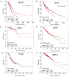
Survival analysis of the ACHE gene in lung adenocarcinoma (LUAD), lung squamous cell carcinoma (LUSC), rectum adenocarcinoma (READ), liver hepatocellular carcinoma (LIHC), stomach adenocarcinoma (STAD), and invasive breast carcinoma (BRCA). (A) Survival analysis for the ACHE gene in LUAD [81]. (B) Survival analysis for the ACHE gene in LUSC [81]. (C) Survival analysis for the ACHE gene in READ [81]. (D) Survival analysis for the ACHE gene in LIHC [81]. (E) Survival analysis for the ACHE gene in STAD [81]. (F) Survival analysis for the ACHE gene in BRCA [81].
Figure 8
Figure 8
Participation of acetylcholinesterase in apoptosome formation. Acetylcholinesterase binds to caveolin-1, which allows the binding of Apaf-1 to cytochrome C, which consequently causes cleavage of procaspase-9 to produce the active form. Then, caspase 9 promotes the activation of the caspase cascade and apoptosis. Created with Biorender.com (accessed on 12 June 2023).
Figure 9
Figure 9
The role of AChE in tumorigenicity. AChE is an essential protein in regulating the ACh levels in non-cholinergic organs, since this neurotransmitter promotes cell proliferation, inhibition of apoptosis, migration, and invasion through acetylcholine receptors (AChRs). (A) In normal conditions, AChE controls ACh levels by hydrolyzing this neurotransmitter, preventing the ACh from reaching AChRs; therefore, the tumor growth is controlled, decreasing tumorigenicity and proliferation. (B) When AChE is chemically inhibited (the BW248c51 inhibitor is impermeable to the cell membrane) or the AChE activity decreases for another cause, the ACh levels are not modified; therefore, this neurotransmitter reaches AChRs, promoting cell proliferation, inhibiting apoptosis, and increasing migration and invasion. This is reflected in tumor growth, increasing tumorigenicity, proliferation, and metastasis. Created with Biorender.com (accessed on 12 June 2023).
Figure 10
Figure 10
The participation of the PI3K/Akt pathway in the deregulation of AChE. Activation of the RTK receptors, nAChRs, and mAChRs could trigger the PI3K/Akt pathway, decreasing AChE content and AChE activity. The low AChE activity could increase ACh levels that stimulate nAChRs and mAChRs, increasing a sustained hyperactivation of the PI3k/Akt and MAP kinase pathways that promote cell proliferation, survival, and sustained AChE decrement. The AchE decrement could affect the apoptosome formation, increasing survival. Created with Biorender.com (accessed on 12 June 2023).
Figure 11
Figure 11
AChE activity regulates cell proliferation in HCC. (A) High AChE activity hydrolyzes to ACh, preventing nAChR and mAChR stimulation. These inactive MAPK and Akt pathways cause a reduction in the GSK3β phosphorylation levels, resulting in greater GSK3β activation, β-catenin degradation, and downregulation of cyclin D1. (B) A drop in AChE activity promotes high ACh levels, stimulating nAChRs and mAChRs. This induces a triggering of the MAPK and Akt pathways, promoting an increment in the GSK3β phosphorylation levels, resulting in the inactivation of GSK3β, which allows reaching a stable level of β-catenin in the cytosol and translocating to the nucleus. This induces upregulation of cyclin D1. The Akt pathway can also decrease both the AChE content and AChE activity. Created with Biorender.com (accessed on 12 June 2023).
Figure 12
Figure 12
Role of AChE from tumor initiation to progression to metastasis. A balance between ACh levels and AChE activity in normal tissue favors tissue homeostasis. During the initial stages of the tumor, AChE levels increase, causing a decrement in ACh concentrations that affect the tumor microenvironment. This allows both the inflammation and TAMs to increase, favoring tumor development. As the MAPK and Akt pathways are hyperactivated, they induce a decrease in AChE that is also potentiated by DNMT1-mediated hypermethylation in aggressive tumors. A reduction in AChE activity increases ACh levels, directly affecting the tumor and inducing cell proliferation, survival, and metastasis. TAMs: tumor-associated macrophages. Created with Biorender.com (accessed on 12 June 2023).

Similar articles

Cited by

References

    1. Horiuchi Y., Kimura R., Kato N., Fujii T., Seki M., Endo T., Kato T., Kawashima K. Evolutional Study on Acetylcholine Expression. Life Sci. 2003;72:1745–1756. doi: 10.1016/S0024-3205(02)02478-5. - DOI - PubMed
    1. Pérez-Aguilar B., Vidal C.J., Gomez-Olivares J.L., Gerardo-Ramirez M., Gutiérrez-Ruiz M.C., Gomez-Quiroz L.E. Acetylcholinesterase. In: Choi S., editor. Encyclopedia of Signaling Molecules. Springer International Publishing; Cham, Switzerland: 2018. pp. 91–101.
    1. Resende R.R., Alves A.S., Britto L.R.G., Ulrich H. Role of Acetylcholine Receptors in Proliferation and Differentiation of P19 Embryonal Carcinoma Cells. Exp. Cell Res. 2008;314:1429–1443. doi: 10.1016/j.yexcr.2008.01.003. - DOI - PubMed
    1. Yu H., Xia H., Tang Q., Xu H., Wei G., Chen Y., Dai X., Gong Q., Bi F. Acetylcholine Acts through M3 Muscarinic Receptor to Activate the EGFR Signaling and Promotes Gastric Cancer Cell Proliferation. Sci. Rep. 2017;7:40802. doi: 10.1038/srep40802. - DOI - PMC - PubMed
    1. Cheng K., Samimi R., Xie G., Shant J., Drachenberg C., Wade M., Davis R.J., Nomikos G., Raufman J.-P. Acetylcholine Release by Human Colon Cancer Cells Mediates Autocrine Stimulation of Cell Proliferation. Am. J. Physiol. Gastrointest. Liver Physiol. 2008;295:G591–G597. doi: 10.1152/ajpgi.00055.2008. - DOI - PMC - PubMed

LinkOut - more resources