. 2023 Sep 14;12(9):1447.
doi: 10.3390/antibiotics12091447.

Pseudouridine Synthase RsuA Confers a Survival Advantage to Bacteria under Streptomycin Stress

Affiliations

Pseudouridine Synthase RsuA Confers a Survival Advantage to Bacteria under Streptomycin Stress

Sudeshi M Abedeera et al. Antibiotics (Basel). .

Abstract

Bacterial ribosome small subunit rRNA (16S rRNA) contains 11 nucleotide modifications scattered throughout all its domains. The 16S rRNA pseudouridylation enzyme, RsuA, which modifies U516, is a survival protein essential for bacterial survival under stress conditions. A comparison of the growth curves of wildtype and RsuA knock-out E. coli strains illustrates that RsuA renders a survival advantage to bacteria under streptomycin stress. The RsuA-dependent growth advantage for bacteria was found to be dependent on its pseudouridylation activity. In addition, the role of RsuA as a trans-acting factor during ribosome biogenesis may also play a role in bacterial growth under streptomycin stress. Furthermore, circular dichroism spectroscopy measurements and RNase footprinting studies have demonstrated that pseudouridine at position 516 influences helix 18 structure, folding, and streptomycin binding. This study exemplifies the importance of bacterial rRNA modification enzymes during environmental stress.

Keywords: helix18; pseudouridine; pseudouridine synthase; ribosome; streptomycin.


Conflict of interest statement

The authors declare no conflict of interest. The funders had no role in the design of the study; in the collection, analyses, or interpretation of data; in the writing of the manuscript; or in the decision to publish the results.

Figures

Figure 1
Figure 1
Streptomycin binds close to 16S helix 18. (a) 16S helix 18 model RNAs used in this research are shown. (b) Streptomycin binds to the pocket formed by ribosomal protein uS12 (yellow) and 16S helices 1 (green), 18 (blue), 27 (cyan), and 44 (pink) as observed in the X-ray crystal structure of streptomycin-bound 30S ribosome (PDB ID 4V50). Blue and red spheres represent Ψ516 and streptomycin, respectively.
Figure 2
Figure 2
RsuA increases the resistance toward streptomycin. The normalized cell growth for Wt (black squares) and ΔRsuA (red squares) E. coli strains are plotted at varying streptomycin concentrations (0–200 μg/mL). The inset shows the corresponding IC50 values (μg/mL) for Wt and ΔRsuA E. coli strains.
Figure 3
Figure 3
RsuA influences bacterial growth kinetics during streptomycin stress. Bacterial growth curves obtained for (a) wildtype (Wt) E. coli and (b) RsuA knock-out (ΔRsuA) strain of E. coli, and RsuA knock-out strain expressing (c) wildtype RsuA (ΔRsuA + wtRsuA) and (d) mutant RsuA (ΔRsuA + mutRsuA) in the background (50 μM IPTG), at varying streptomycin concentrations from 0–13.5 μg/mL, are shown. The average of the biological triplicates is shown.
Figure 4
Figure 4
The 17S/16S ratio for wildtype (Wt) and RsuA knock-out (ΔRsuA) strains of E. coli in the presence and absence of streptomycin (Sm) stress. Error bars represent the SD of triplicates.
Figure 5
Figure 5
The Ψ516 modification influences the folding of helix 18. (a) The 16S helix 18 in the 30S X-ray crystal structure (PDB ID: 4V50) is shown. Blue spheres represent Mg2+ present near helix 18. Ψ516 is shown in magenta. (b) A comparison of circular dichroism (CD) spectra for h18-Ψ (green) and h18-U (blue) model RNAs. CD spectra of (c) h18-Ψ and (d) h18-U at various magnesium concentrations are shown. The inset shows the change in the 250–270 nm wavelength region of each spectrum. Changes in molar ellipticity with [Mg2+] for (e) h18-Ψ and (f) h18-U are shown. Standard deviation of triplicates is shown as error bars.
Figure 6
Figure 6
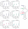
Ψ516 modification changes local RNA structure. (a) Radiograph showing RNase T1 digestion pattern of h18-Ψ (left panel) and h18-U (right panel) model RNAs. Bands corresponding to RNase T1 cleavages at G524, G521, G517, and G515 are shown in blue, green, purple, and teal arrows, respectively. The relative cleavage at each guanine of h18-Ψ (b,c) and h18-U (dg) compared with full-length intact RNA at various Mg2+ concentrations (mM) are plotted.
Figure 7
Figure 7
Ψ516 modification increases the affinity of streptomycin to helix 18. (a) CD spectra obtained for the h18-Ψ model RNA, at various streptomycin concentrations (0–3 mM) are shown. Experiments were performed at 30 °C in CD buffer (20 mM potassium cacodylate pH 7.0, 15 mM KCl, 4 mM MgCl2). (b) The fraction of RNA complexed with streptomycin at each streptomycin concentration is shown. The dissociation constants (Kds) of streptomycin binding for h18-Ψ and h18-U model RNAs were obtained by least-square fitting of binding curves to binding isotherm. All the experiments were undertaken in triplicate to confirm the reproducibility.
Figure 8
Figure 8
Streptomycin interacts with different regions of the h18 model RNAs with contrasting affinities. Relative cleavage at (a) G524 and (b) G521 of h18-Ψ and (c) G524, (d) G521, (e) G517, and (f) G515 of h18-U model RNAs at various concentrations of streptomycin are shown. Error bars shown on graphs represent the standard deviation of three replicates.

References

    1. Chow C.S., Lamichhane T.N., Mahto S.K. Expanding the Nucleotide Repertoire of the Ribosome with Post-Transcriptional Modifications. ACS Chem. Biol. 2007;2:610–619. doi: 10.1021/cb7001494. - DOI - PMC - PubMed
    1. Helm M. Post-transcriptional nucleotide modification and alternative folding of RNA. Nucleic Acids Res. 2006;34:721–733. doi: 10.1093/nar/gkj471. - DOI - PMC - PubMed
    1. Douthwaite S., Fourmy D., Yoshizawa S. In: Fine-Tuning of RNA Functions by Modification and Editing. 2nd ed. Grosjean H., editor. Volume 12. Springer; Berlin/Heidelberg, Germany: 2005. pp. 285–307.
    1. Lázaro E., Rodriguez-Fonseca C., Porse B., Ureña D., Garrett R.A., Ballesta J.P.G. A Sparsomycin-resistant Mutant of Halobacterium salinarium Lacks a Modification at Nucleotide U2603 in the Peptidyl Transferase Centre of 23 S rRNA. J. Mol. Biol. 1996;261:231–238. doi: 10.1006/jmbi.1996.0455. - DOI - PubMed
    1. Toh S.M., Mankin A.S. An indigenous post-transcriptional modification in the ribosomal peptidyl transferase center confers resistance to an array of protein synthesis inhibitors. J. Mol. Biol. 2008;380:593–597. doi: 10.1016/j.jmb.2008.05.027. - DOI - PMC - PubMed

LinkOut - more resources