. 2023 Aug 22;11(9):2336.
doi: 10.3390/biomedicines11092336.

Morphofunctional Changes in Brain and Peripheral Blood in Adult and Aged Wistar Rats with AlCl3-Induced Neurodegeneration

Affiliations

Morphofunctional Changes in Brain and Peripheral Blood in Adult and Aged Wistar Rats with AlCl3-Induced Neurodegeneration

Alexandra Vladislavovna Sentyabreva et al. Biomedicines. .

Abstract

Background: the general lifespan has been prolonged greatly during the past century, and the incidence of age-associated diseases, including neurodegenerative ones, has increased as well. However, modelling of age-related pathologies is mostly conducted on adult rodents. We studied morphofunctional changes in the brain and peripheral blood of adult Wistar rats in comparison with old Wistar rats to determine age-related physiological changes and differences in adaptive reactions to AlCl3 exposure.

Methods: the work was performed on adult and old male Wistar rats. The animals consumed a 100 mg/kg solution of AlCl3 each day for 60 days. Morphological changes of neurons and microglia, mRNA expression levels of pro-inflammatory and anti-inflammatory cytokines, microglia activation markers, amyloid-related proteins, and hallmarks of cellular senescence, monocyte, and lymphocyte subpopulations in the peripheral blood were examined.

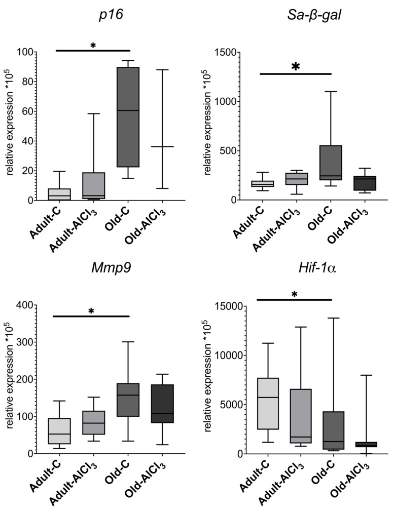
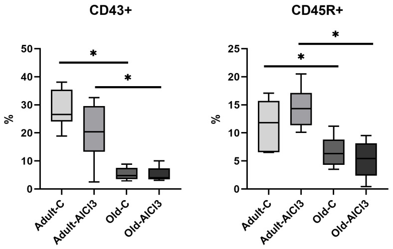
Results: old rats showed increasing hyperchromic neurons in the hippocampus; activation of microglia; upregulation of pro-inflammatory cytokines and cellular senescence markers; downregulation of anti-inflammatory cytokines; and Hif-1a and a decrease in B-cells and monocyte in peripheral blood.

Conclusion: compared to young animals, aged rats respond to aluminum exposure with a severe decline of most cells' function and irreversible neuronal loss. Regarding all reported data, neurodegeneration modelling and investigating of factors capable of accelerating or preventing it should be performed in experimental work on aged animals.

Keywords: Alzheimer’s disease; aging; animal models; inflammaging; inflammation; neurodegeneration.


Conflict of interest statement

The authors declare no conflict of interest.

Figures

Figure 1
Figure 1
Time (s) required to reach the platform during acquisition trials for adult and old Wistar rats of control and experimental groups on day 0 and day 56 of the experiment. Adult-C 0—adult rats control group (n = 8), the trial on day 0 of the experiment; Adult-C 56—adult rats control group (n = 8), the trial on day 56 of the experiment; Adult-AlCl3 0—adult rats that consumed AlCl3 (n = 8), the trial on day 0 of the experiment; Adult-AlCl3 56—adult rats that consumed AlCl3 (n = 8), the trial on day 56 of the experiment; Old-C 0—old rats control group (n = 8), the trial on day 0 of the experiment; Old-C 56—old rats control group (n = 8), the trial on day 56 of the experiment; Old-AlCl3 0—old rats that consumed AlCl3 (n = 8), the trial on day 0 of the experiment; Old-AlCl3 56—old rats that consumed AlCl3 (n = 8), the trial on day 56 of the experiment. The data displayed as: line—median, box—25–75 quartiles, whiskers—nonoutlier range; *—p < 0.05. The Kruskal–Wallis test was used for multiple comparisons.
Figure 2
Figure 2
CA1 (A,C,E,G) and CA3 (B,D,F,H) zones of the hippocampus in rats of Adult-C group (A,B), Adult-AlCl3 group (C,D), Old-C group (E,F), and Old-AlCl3 group (G,H). Nissl’s staining, ×400.
Figure 2
Figure 2
CA1 (A,C,E,G) and CA3 (B,D,F,H) zones of the hippocampus in rats of Adult-C group (A,B), Adult-AlCl3 group (C,D), Old-C group (E,F), and Old-AlCl3 group (G,H). Nissl’s staining, ×400.
Figure 3
Figure 3
The percentage of hyperchromic neurons in zones CA1, CA3, and in the dentate gyrus of the hippocampus in adult and old Wistar rats of control and experimental groups. HN—hyperchromic neurons; Adult-C—adult rats control group (n = 8); Adult-AlCl3—adult rats that consumed AlCl3 (n = 8); Old-C—old rats control group (n = 8); Old-AlCl3—old rats that consumed AlCl3 (n = 8). The data displayed as: line—median, box—25–75 quartiles, whiskers—nonoutlier range; *—p < 0.05. The Kruskal–Wallis test was used for multiple comparisons.
Figure 4
Figure 4
Morphological characteristics of microglia cells with thin and short processes in both Adult-C (A) and Adult-AlCl3 (B) groups; enlarged microglia with spheroidal swelling, hypertrophic, beaded, and tortuous processes in Old-C rats (C); and microglia of an increased size with beaded, tortuous, and fragmented but not thickened processes in Old-AlCl3 rats (D). Iba-1 antibody + HRP secondary antibody IHC and hematoxylin staining, ×1600. Adult-C—adult rats control group (n = 8); Adult-AlCl3—adult rats that consumed AlCl3 (n = 8); Old-C—old rats control group (n = 8); Old-AlCl3—old rats that consumed AlCl3 (n = 8).
Figure 5
Figure 5
Mrna expression levels of Il-18 and Tnf-α in the prefrontal cortex of adult and old Wistar rats in control and experimental groups. Adult-C—adult rats control group (n = 7); Adult-AlCl3—adult rats that consumed AlCl3 (n = 8); Old-C—old rats control group (n = 8) Old-AlCl3—old rats that consumed AlCl3 (n = 8). The data displayed as: line—median, box—25–75 quartiles, whiskers—nonoutlier range; *—p < 0.05. The Kruskal–Wallis test was used for multiple comparisons.
Figure 6
Figure 6
mRNA expression levels of Il-10 and Tgf-β in the prefrontal cortex of adult and old Wistar rats in control and experimental groups. Adult-C—adult rats control group (n = 7); Adult-AlCl3—adult rats that consumed AlCl3 (n = 8); Old-C—old rats control group (n = 8); Old-AlCl3—old rats that consumed AlCl3 (n = 8). The data displayed as: line—median, box—25–75 quartiles, whiskers—nonoutlier range; *—p < 0.05. The Kruskal–Wallis test was used for multiple comparisons.
Figure 7
Figure 7
mRNA expression levels of iNos and Cd163+ and M1/M2 (iNos/Cd163+) ratio in the prefrontal cortex in adult and old Wistar rats of control and experimental groups. Adult-C—adult rats control group (n = 7); Adult-AlCl3—adult rats that consumed AlCl3 (n = 8); Old-C—old rats control group (n = 6); Old-AlCl3—old rats that consumed AlCl3 (n = 5); The data displayed as: line—median, box—25–75 quartiles, whiskers—nonoutlier range; *—p < 0.05. The Kruskal–Wallis test was used for multiple comparisons.
Figure 8
Figure 8
mRNA expression levels in App and Bace1 in the prefrontal cortex of adult and old Wistar rats of control and experimental groups. Adult-C—adult rats control group (n = 7); Adult-AlCl3—adult rats that consumed AlCl3 (n = 8); Old-C—old rats control group (n = 8); Old-AlCl3—old rats that consumed AlCl3 (n = 8); The data displayed as: line—median, box—25–75 quartiles, whiskers—nonoutlier range; *—p < 0.05. The Kruskal–Wallis test was used for multiple comparisons.
Figure 9
Figure 9
mRNA expression levels of p16, Sa-β-gal, Mmp9, and Hif-1α in the prefrontal cortex in adult and old Wistar rats of control and experimental groups. Adult-C—adult rats control group (n = 5); Adult-AlCl3—adult rats that consumed AlCl3 (n = 6); Old-C—old rats control group (n = 8); Old-AlCl3—old rats that consumed AlCl3 (n = 5); The data displayed as: line—median, box—25–75 quartiles, whiskers—nonoutlier range; *—p < 0.05. The Kruskal–Wallis test was used for multiple comparisons.
Figure 10
Figure 10
The percentage of CD45R+ B-cells and CD43+ monocyte in peripheral blood of adult and old Wistar rats of control and experimental groups. Adult-C—adult rats control group (n = 8); Adult-AlCl3—adult rats that consumed AlCl3 (n = 8); Old-C—old rats control group (n = 8); Old-AlCl3—old rats that consumed AlCl3 (n = 8). The data displayed as: line—median, box—25–75 quartiles, whiskers—nonoutlier range; *—p < 0.05. The Kruskal–Wallis test was used for multiple comparisons.
Figure 11
Figure 11
Morphofunctional changes in brain and peripheral blood in adult and old Wistar rats due to aging and neurodegeneration modelling. Adult-C—adult rats control group, Old-C—old rats control group, Adult-AlCl3—adult rats that consumed AlCl3, Old-AlCl3—old rats that consumed AlCl3, DG—dentate gyrus, TNF-α—tumor necrosis factor α, IL-18—interleukin 18, IL10—interleukin 10, TGF-β—transforming growth factor β, iNOS—inducible nitric oxide synthase, M1/M2 ratio—ratio of mRNA expression of markers for M1-type activated microglia to M2-type activated microglia, MMP9—matrix metallopeptidase 9, p16—cyclin-dependent kinase inhibitor 2A, Sa-β-gal—senescence-associated-β-galactosidase, HIF-1α—hypoxia-inducible factor 1α, APP—amyloid precursor protein, BACE1—β-Site APP-cleaving enzyme 1.

Similar articles

Cited by

References

    1. WHO Dementia. 2021. [(accessed on 19 June 2023)]. Available online: https://www.who.int/publications/i/item/9789241550543.
    1. Streit W.J., Braak H., Del Tredici K., Leyh J., Lier J., Khoshbouei H., Eisenlöffel C., Müller W., Bechmann I. Microglial Activation Occurs Late During Preclinical Alzheimer’s Disease. Glia. 2018;66:2550–2562. doi: 10.1002/glia.23510. - DOI - PubMed
    1. van Dyck C.H., Swanson C.J., Aisen P., Bateman R.J., Chen C., Gee M., Kanekiyo M., Li D., Reyderman L., Cohen S., et al. Lecanemab in Early Alzheimer’s Disease. N. Engl. J. Med. 2023;388:9–21. doi: 10.1056/NEJMoa2212948. - DOI - PubMed
    1. Franceschi C., Campisi J. Chronic Inflammation (Inflammaging) and Its Potential Contribution to Age-Associated Diseases. J. Gerontol. A Ser. Biol. Sci. Med. Sci. 2014;69((Suppl. S1)):S4–S9. doi: 10.1093/gerona/glu057. - DOI - PubMed
    1. Dafsari F.S., Jessen F. Depression—An underrecognized target for prevention of dementia in Alzheimer’s disease. Transl. Psychiatry. 2020;10:160. doi: 10.1038/s41398-020-0839-1. - DOI - PMC - PubMed

LinkOut - more resources