. 2023 Aug 23;11(9):2350.
doi: 10.3390/biomedicines11092350.

Is Spheroid a Relevant Model to Address Fibrogenesis in Keloid Research?

Affiliations

Is Spheroid a Relevant Model to Address Fibrogenesis in Keloid Research?

Zélie Dirand et al. Biomedicines. .

Abstract

Keloid refers to a fibro-proliferative disorder characterized by an accumulation of extracellular matrix at the dermis level, overgrowing beyond the initial wound and forming tumor-like nodule areas. The absence of treatment for keloid is clearly related to limited knowledge about keloid etiology. In vitro, keloids were classically studied through fibroblasts monolayer culture, far from keloid in vivo complexity. Today, cell aggregates cultured as 3D spheroid have gained in popularity as new tools to mimic tissue in vitro. However, no previously published works on spheroids have specifically focused on keloids yet. Thus, we hypothesized that spheroids made of keloid fibroblasts (KFs) could be used to model fibrogenesis in vitro. Our objective was to qualify spheroids made from KFs and cultured in a basal or pro-fibrotic environment (+TGF-β1). As major parameters for fibrogenesis assessment, we evaluated apoptosis, myofibroblast differentiation and response to TGF-β1, extracellular matrix (ECM) synthesis, and ECM-related genes regulation in KFs spheroids. We surprisingly observed that fibrogenic features of KFs are strongly downregulated when cells are cultured in 3D. In conclusion, we believe that spheroid is not the most appropriate model to address fibrogenesis in keloid, but it constitutes an efficient model to study the deactivation of fibrotic cells.

Keywords: ECM; TGF-β1; fibrosis; keloid fibroblast; spheroid; α-SMA.


Conflict of interest statement

The authors declare no conflict of interest.

Figures

Figure 1
Figure 1
KFs and NDFs area equally evolve over time independently from TGF-β1 activation. To generate spheroids, NDFs or KFs were seeded in ULA culture plates +/− TGF-β1. (A) Diameters were followed over time using IncuCyteS3 microscope. (B) After maturation (7 days), spheroids slices were stained with H&E for architecture study. Results are expressed as mean ± SD. Statistical analyses were performed using two-way ANOVA (n= 16 spheroids per condition).
Figure 2
Figure 2
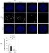
TGF-β1 reduced apoptotic cells rate in KFs spheroids more than in NDFs ones. (A) After 7 days, NDFs and KFs spheroids (either culture in control medium or with 10 ng/mL TGF-β1) were prepared for TUNEL staining (7 µm spheroid section). Images from confocal microscope show total nuclei in blue and those from apoptotic cells in red (highlighted by the white arrows). (B) TUNEL positive cells were counted on spheroid sections. Results are represented as a number of positive cells/mm2. A minimum of five slides were used for each spheroid [n = 4 spheroid per condition]. Statistical analyses were performed using two-way ANOVA * for p < 0.05.
Figure 3
Figure 3
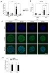
TGF-β1-induced α-SMA expression discontinues in KFs spheroids. α-SMA expression was evaluated in NDFs and KFs monolayers and spheroids after 7 days of treatment with or without TGF-β1. The expression of α-SMA was studied using ELISA (A) and RT-qPCR (B). After maturation, spheroids thus treated were immunostained for α-SMA observation (C) and semi quantification (D) [n = 10 spheroid and n= 2–6 images per spheroid]. Statistical analyses were performed using two-way ANOVA * for p < 0.05; ** for p < 0.005; *** for p < 0.001.
Figure 4
Figure 4
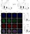
Three-Dimensional culture and TGF-β1 activation converge to downregulate CD26 and TGFβRII expression. TGFβRII (A) and DPP4 (B) mRNA synthesis were evaluated by RT-qPCR in monolayers and spheroids performed either with NDFs or KFs in the presence of TGF-β1 vs. control [n = 4 per condition]. After 7 days of treatment, spheroids thus treated were immunostained for CD26 (green) and TGFβRII (red) observation (C) and semi quantification (D,E). Nuclei were counterstained with DAPI (Blue). [n = 10 spheroid and n= 2–6 images per spheroid]. Statistical analyses were performed using two-way ANOVA * for p < 0.05; ** for p < 0.005; *** for p < 0.001; **** for p < 0.0001.
Figure 5
Figure 5
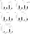
Overexpression of ECM related genes discontinues when KFs are cultured from 2D to 3D. Fibronectin expression was assessed and quantified using ELISA (A) and RT-qPCR (B) in monolayers and spheroids made with NDFs and KFs in the presence of TGF-β1 vs. control. Type I (C) and type III (D) collagen encoding mRNA, and COL1A1/COL3A1 ratio (E) were evaluated by RT-qPCR in monolayers or spheroids produced and treated as previously described [n = 4 spheroids per condition]. Statistical analyses were performed using two-way ANOVA * for p < 0.05; ** for p < 0.005; *** for p < 0.001; **** for p < 0.0001.

References

    1. WHO ICD-11 for Mortality and Morbidity Statistics. [(accessed on 27 July 2023)]; Available online: https://icd.who.int/browse11/l-m/en#/http%3a%2f%2fid.who.int%2ficd%2fent....
    1. Ogawa R. Keloid and Hypertrophic Scars Are the Result of Chronic Inflammation in the Reticular Dermis. Int. J. Mol. Sci. 2017;18:606. doi: 10.3390/ijms18030606. - DOI - PMC - PubMed
    1. Tan S., Khumalo N., Bayat A. Understanding Keloid Pathobiology from a Quasi-Neoplastic Perspective: Less of a Scar and More of a Chronic Inflammatory Disease with Cancer-Like Tendencies. Front. Immunol. 2019;10:1810. doi: 10.3389/fimmu.2019.01810. - DOI - PMC - PubMed
    1. Betarbet U., Blalock T.W. Keloids: A Review of Etiology, Prevention, and Treatment. J. Clin. Aesthet. Dermatol. 2020;13:33–43. - PMC - PubMed
    1. Supp D.M. Animal Models for Studies of Keloid Scarring. Adv. Wound Care. 2019;8:77–89. doi: 10.1089/wound.2018.0828. - DOI - PMC - PubMed