. 2023 Aug 31;11(9):2439.
doi: 10.3390/biomedicines11092439.

Acoustic Voice Analysis as a Useful Tool to Discriminate Different ALS Phenotypes

Affiliations

Acoustic Voice Analysis as a Useful Tool to Discriminate Different ALS Phenotypes

Giammarco Milella et al. Biomedicines. .

Abstract

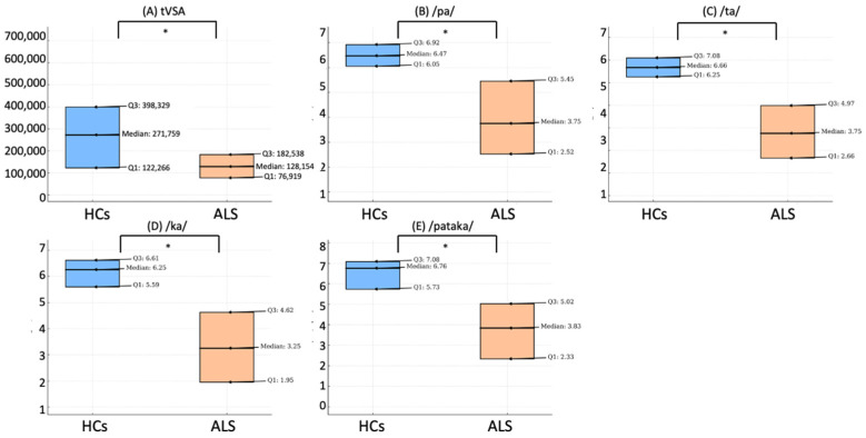
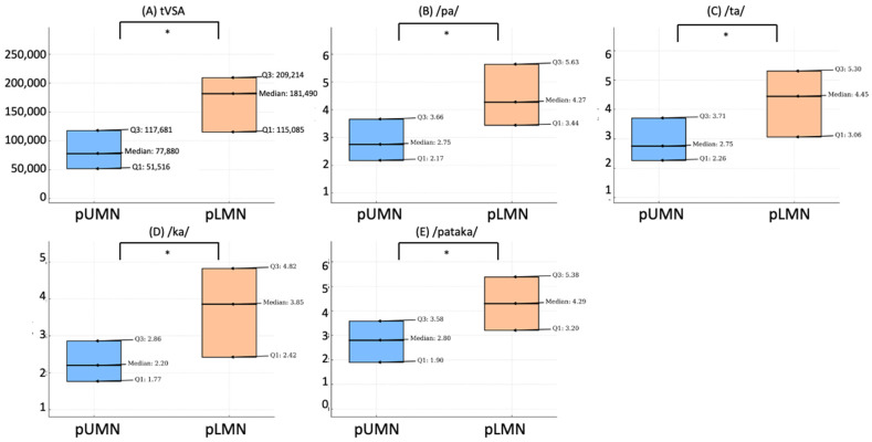
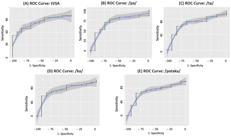
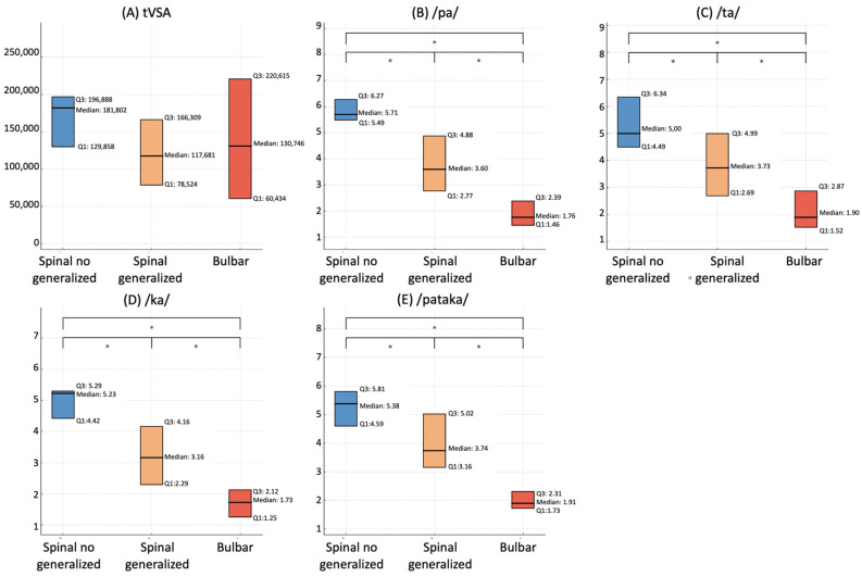
Approximately 80-96% of people with amyotrophic lateral sclerosis (ALS) become unable to speak during the disease progression. Assessing upper and lower motor neuron impairment in bulbar regions of ALS patients remains challenging, particularly in distinguishing spastic and flaccid dysarthria. This study aimed to evaluate acoustic voice parameters as useful biomarkers to discriminate ALS clinical phenotypes. Triangular vowel space area (tVSA), alternating motion rates (AMRs), and sequential motion rates (SMRs) were analyzed in 36 ALS patients and 20 sex/age-matched healthy controls (HCs). tVSA, AMR, and SMR values significantly differed between ALS and HCs, and between ALS with prevalent upper (pUMN) and lower motor neuron (pLMN) impairment. tVSA showed higher accuracy in discriminating pUMN from pLMN patients. AMR and SMR were significantly lower in patients with bulbar onset than those with spinal onset, both with and without bulbar symptoms. Furthermore, these values were also lower in patients with spinal onset associated with bulbar symptoms than in those with spinal onset alone. Additionally, AMR and SMR values correlated with the degree of dysphagia. Acoustic voice analysis may be considered a useful prognostic tool to differentiate spastic and flaccid dysarthria and to assess the degree of bulbar involvement in ALS.

Keywords: ALS; ALS phenotypes; bulbar impairment; voice analysis.


Conflict of interest statement

The authors report there are no competing interest to declare.

Figures

Figure 1
Figure 1
Comparison of voice parameters between healthy controls and ALS patients. This figure illustrates the differences in various voice parameters between healthy controls (HCs) and patients with amyotrophic lateral sclerosis (ALS). Box plot of tVSA (A); articulation rate of the syllable /pa/ (B); /ta/ (C); /ka/ (D); and the sequence /pataka/ (E). Each plot illustrates the median, first quartile (Q1), and third quartile (Q3) values, comparing the distributions between HCs and ALS patients. The symbol * denotes a significant difference at p < 0.05.
Figure 2
Figure 2
Comparison of voice parameters between prevalent UMN and LMN ALS patients. This figure shows the differences in voice parameters between ALS patients with prevalent upper motor neuron (pUMN) and prevalent lower motor neuron (pLMN). Box plot of tVSA (A); articulation rate of the syllable /pa/ (B); /ta/ (C); /ka/ (D); and the sequence /pataka/ (E). Each plot illustrates the median, first quartile (Q1), and third quartile (Q3) values, comparing the distributions between these two ALS phenotypes. The symbol * denotes a significant difference at p < 0.05.
Figure 3
Figure 3
Discrimination of ALS patients with prevalent UMN and LMN impairment using voice parameters. The figure shows the ROC curves for various voice parameters able to discriminate ALS patients with prevalent upper motor neuron (pUMN) impairment and those with prevalent lower motor neuron (pLMN) impairment. ROC curve of tVSA (A); articulation rate of the syllable /pa/ (B); /ta/ (C); /ka/ (D); and the sequence /pataka/ (E). The area under each curve quantifies the accuracy of the respective voice parameter as a diagnostic tool for distinguishing these two ALS phenotypes.
Figure 4
Figure 4
Comparison of voice parameters according to the site of onset in ALS patients. This figure illustrates the differences in various voice parameters among ALS patients, grouped according to their site of disease onset. Box plot of tVSA (A); articulation rate of the syllable /pa/ (B); /ta/ (C); /ka/ (D); and the sequence /pataka/ (E). Each plot illustrates the median, first quartile (Q1), and third quartile (Q3) values, comparing the distributions between patients with spinal onset (both with and without evidence of bulbar symptoms at clinical evaluation) and those with bulbar onset. The symbol * denotes a significant difference at p < 0.05.

Similar articles

Cited by

References

    1. Kiernan M.C., Vucic S., Cheah B.C., Turner M.R., Eisen A., Hardiman O., Burrell J.R., Zoing M.C. Amyotrophic lateral sclerosis. Lancet. 2011;377:942–955. doi: 10.1016/S0140-6736(10)61156-7. - DOI - PubMed
    1. Brooks B.R. El Escorial World Federation of Neurology criteria for the diagnosis of amyotrophic lateral sclerosis. Subcommittee on Motor Neuron Diseases/Amyotrophic Lateral Sclerosis of the World Federation of Neurology Research Group on Neuromuscular Diseases and the El Escorial “Clinical limits of amyotrophic lateral sclerosis” workshop contributors. J. Neurol. Sci. 1994;124:96–107. doi: 10.1016/0022-510x(94)90191-0. - DOI - PubMed
    1. Brooks B.R., Miller R.G., Swash M., Munsat T.L., World Federation of Neurology Research Group on Motor Neuron Diseases El Escorial revisited: Revised criteria for the diagnosis of amyotrophic lateral sclerosis. Amyotroph. Lateral Scler. Other Mot. Neuron Disord. 2000;1:293–299. doi: 10.1080/146608200300079536. - DOI - PubMed
    1. Hannaford A., Pavey N., van den Bos M., Geevasinga N., Menon P., Shefner J.M., Kiernan M.C., Vucic S. Diagnostic Utility of Gold Coast Criteria in Amyotrophic Lateral Sclerosis. Ann. Neurol. 2021;89:979–986. doi: 10.1002/ana.26045. - DOI - PubMed
    1. de Carvalho M., Dengler R., Eisen A., England J.D., Kaji R., Kimura J., Mills K., Mitsumoto H., Nodera H., Shefner J., et al. Electrodiagnostic criteria for diagnosis of ALS. Clin. Neurophysiol. 2008;119:497–503. doi: 10.1016/j.clinph.2007.09.143. - DOI - PubMed

LinkOut - more resources