

Effect of Calcitriol and Vitamin D Receptor Modulator 2 on Recovery of Injured Skeletal Muscle in Wistar Rats
- PMID: 37760917
- PMCID: PMC10525631
- DOI: 10.3390/biomedicines11092477
Effect of Calcitriol and Vitamin D Receptor Modulator 2 on Recovery of Injured Skeletal Muscle in Wistar Rats
Abstract
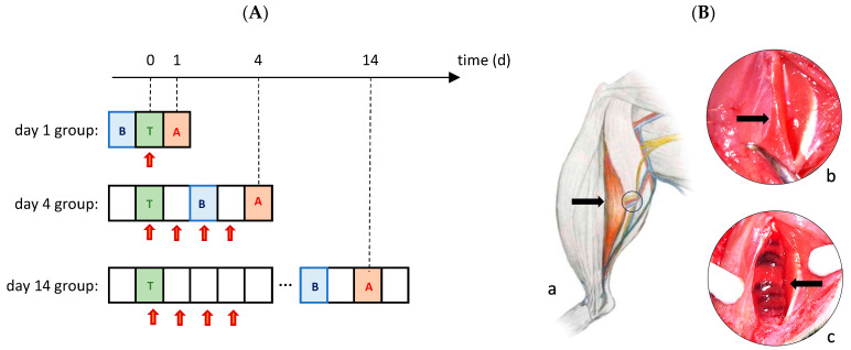
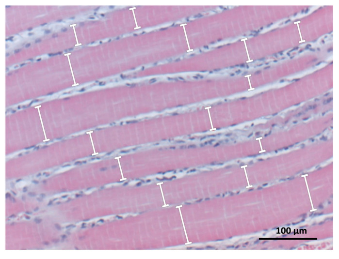
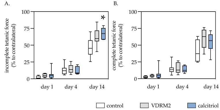
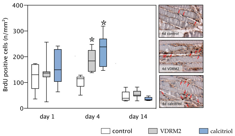
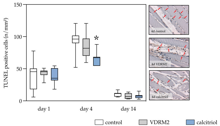
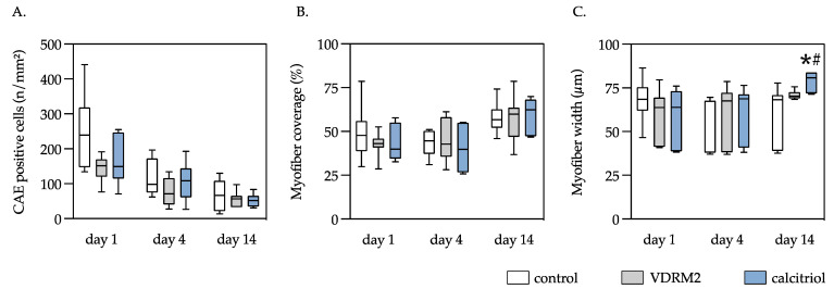
Muscle injuries often result in functional limitations due to insufficient healing. This study assessed the influence of calcitriol and vitamin D Receptor Modulator 2 (VDRM2) on muscle regeneration in male Wistar rats following open blunt muscle injury. The injured left soleus muscle of the rats was treated for the first four days after trauma with local injections of either calcitriol, VDRM2, or a 10% ethanol solution (control). Although muscle strength significantly decreased post-injury, all groups showed gradual improvement but did not achieve full recovery. By the 14th day, calcitriol-treated rats significantly outperformed the control group in the incomplete tetanic force, with VDRM2-treated rats showing muscle strength values that fell between the control and calcitriol groups. Similar trends were observed in complete tetanic contractions and were confirmed histologically via muscle cell width quantification. Additionally, histological analysis showed increased cellular turnover on the fourth postoperative day in the calcitriol group, as indicated by elevated cell proliferation rates and fewer apoptotic cells. VDRM2-treated animals showed only an increased proliferative activity on day 4 after injury. No noticeable differences between the groups for CAE-positive cells or visible muscle tissue area were found. In conclusion, predominantly calcitriol positively influenced post-trauma muscle recovery, where VDRM2 had substantially lower biological activity.
Keywords: calcitriol; cellular turnover; healing; muscle injury; vitamin D receptor modulator 2 (VDRM2).
Conflict of interest statement
The authors declare no conflict of interest.
Figures
Similar articles
-
Vitamin D increases cellular turnover and functionally restores the skeletal muscle after crush injury in rats.Am J Pathol. 2013 Mar;182(3):895-904. doi: 10.1016/j.ajpath.2012.11.006. Epub 2012 Dec 20. Am J Pathol. 2013. PMID: 23260772
-
A nonsecosteroidal vitamin D receptor ligand with improved therapeutic window of bone efficacy over hypercalcemia.J Bone Miner Res. 2010 Jun;25(6):1326-36. doi: 10.1002/jbmr.15. J Bone Miner Res. 2010. PMID: 20200930
-
Granulocyte-colony stimulating factor enhances muscle proliferation and strength following skeletal muscle injury in rats.J Appl Physiol (1985). 2007 Nov;103(5):1857-63. doi: 10.1152/japplphysiol.00066.2007. Epub 2007 Aug 23. J Appl Physiol (1985). 2007. PMID: 17717125
-
Vitamin D, Skeletal Muscle Function and Athletic Performance in Athletes-A Narrative Review.Nutrients. 2019 Aug 4;11(8):1800. doi: 10.3390/nu11081800. Nutrients. 2019. PMID: 31382666 Free PMC article. Review.
-
Vitamin D Promotes Skeletal Muscle Regeneration and Mitochondrial Health.Front Physiol. 2021 Apr 14;12:660498. doi: 10.3389/fphys.2021.660498. eCollection 2021. Front Physiol. 2021. PMID: 33935807 Free PMC article. Review.
Cited by
-
Transcriptomic profiling of skeletal muscle in the DMDmdx rat model of Duchenne muscular dystrophy.Sci Rep. 2025 Aug 11;15(1):29312. doi: 10.1038/s41598-025-14756-9. Sci Rep. 2025. PMID: 40790324 Free PMC article.
-
Vitamin D and Sarcopenia: Implications for Muscle Health.Biomedicines. 2025 Jul 31;13(8):1863. doi: 10.3390/biomedicines13081863. Biomedicines. 2025. PMID: 40868119 Free PMC article. Review.
-
The association between dietary vitamins and the risk of sarcopenia in adults aged 20-59: a study based on the NHANES database.Front Nutr. 2025 Mar 26;12:1535190. doi: 10.3389/fnut.2025.1535190. eCollection 2025. Front Nutr. 2025. PMID: 40206945 Free PMC article.
-
The Importance of Vitamin D and Magnesium in Athletes.Nutrients. 2025 May 13;17(10):1655. doi: 10.3390/nu17101655. Nutrients. 2025. PMID: 40431395 Free PMC article. Review.
References
-
- Owens D.J., Sharples A.P., Polydorou I., Alwan N., Donovan T., Tang J., Fraser W.D., Cooper R.G., Morton J.P., Stewart C., et al. A systems-based investigation into vitamin D and skeletal muscle repair, regeneration, and hypertrophy. Am. J. Physiol.-Endocrinol. Metab. 2015;309:E1019–E1031. doi: 10.1152/ajpendo.00375.2015. - DOI - PubMed
Grants and funding
LinkOut - more resources
Full Text Sources
