Review
. 2023 Sep 13;11(9):2526.
doi: 10.3390/biomedicines11092526.

Cellular and Molecular Processes in Wound Healing

Affiliations
Review

Cellular and Molecular Processes in Wound Healing

Montserrat Fernández-Guarino et al. Biomedicines. .

Abstract

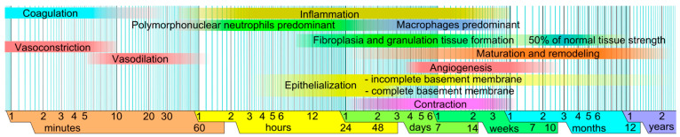
This review summarizes the recent knowledge of the cellular and molecular processes that occur during wound healing. However, these biological mechanisms have yet to be defined in detail; this is demonstrated by the fact that alterations of events to pathological states, such as keloids, consisting of the excessive formation of scars, have consequences yet to be defined in detail. Attention is also dedicated to new therapies proposed for these kinds of pathologies. Awareness of these scientific problems is important for experts of various disciplines who are confronted with these kinds of presentations daily.

Keywords: acute wounds; cellular infiltrate; chronic wounds; keloids; scars.


Conflict of interest statement

The authors declare no conflict of interest.

Figures

Figure 1
Figure 1
Times for the various stages of wound healing on a logarithmic scale, with faded intervals indicating significant variance, primarily based on the size of the lesion and the circumstances surrounding its healing [15].
Figure 2
Figure 2
The main cell types that are involved in the composition of the cellular infiltrate during wound healing: (A) granulocytes stained with Ly6G (B); mast cells stained with avidin; (C) plasmacytoid dendritic cells stained with CD303 (BDCA2); (D) pericytes stained with PDGF. Fluorescence microscopy; scale bar = 10 microns (courtesy of co-author, Stefano Bacci).
Figure 3
Figure 3
The activation of the skin microenvironment in conditions of injury such as a wound. The responses of keratinocytes and cellular infiltrate (see green arrows for respective locations). HE staining and light microscopy; scale bar = 10 microns (courtesy of co-author, Stefano Bacci).
Figure 4
Figure 4
The co-culture of keratinocytes and fibroblasts stained for β-catenin (Alexa red) and collagen type I (Alexa green). (A) DAPI-stained nuclei; (B) Fibroblasts labeled for collagen type I with Alexa green; (C) Keratinocytes labeled for β-catenin with Alexa red; (D) merged image. Confocal microscopy; scale bar = 100 μm (courtesy of co-author, Maria Luisa Hernández-Bule).

Similar articles

Cited by

References

    1. Han G., Ceilley R. Chronic Wound Healing: A Review of Current Management and Treatments. Adv. Ther. 2017;34:599–610. doi: 10.1007/s12325-017-0478-y. - DOI - PMC - PubMed
    1. Sen C.K. Human Wounds and Its Burden: An Updated Compendium of Estimates. Adv. Wound Care. 2019;8:39–48. doi: 10.1089/wound.2019.0946. - DOI - PMC - PubMed
    1. Bacci S. Cellular Mechanisms and Therapies in Wound Healing: Looking toward the Future. Biomedicines. 2021;9:1611. doi: 10.3390/biomedicines9111611. - DOI - PMC - PubMed
    1. Gupta S., Andersen C., Black J., Fife C., Lantis J.I., Niezgoda J., Snyder R., Sumpio B., Tettelbach W., Treadwell T., et al. Management of Chronic Wounds: Diagnosis, Preparation, Treatment, and Follow-up. Wounds Compend. Clin. Res. Pract. 2017;29:S19–S36. - PubMed
    1. Babalska Z., Korbecka-Paczkowska M., Karpiński T.M. Wound Antiseptics and European Guidelines for Antiseptic Application in Wound Treatment. Pharmaceuticals. 2021;14:1253. doi: 10.3390/ph14121253. - DOI - PMC - PubMed

LinkOut - more resources