

Retinal Microvascular Alterations in a Patient with Type 1 Diabetes Mellitus, Hemoglobin D Hemoglobinopathy, and High Myopia-Case Report and Review of the Literature
- PMID: 37761301
- PMCID: PMC10527753
- DOI: 10.3390/diagnostics13182934
Retinal Microvascular Alterations in a Patient with Type 1 Diabetes Mellitus, Hemoglobin D Hemoglobinopathy, and High Myopia-Case Report and Review of the Literature
Abstract
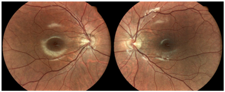
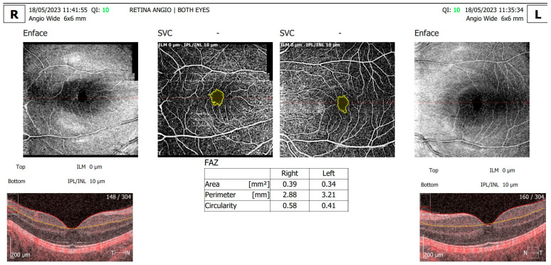
Type 1 diabetes mellitus (type 1 DM) is one of the most prevalent endocrinological diseases among children and young adults, with a growing incidence rate reaching up to 2.9 new cases per year per 100,000 persons below 15 years of age. We report a rare case of a 20-year-old female patient with type 1 DM, hemoglobin D (HbD) heterozygote variant and high myopia of -10.00 spheric diopters, and describe the retinal microvascular alterations visible on OCT angiography (angio-OCT). The patient also presented with a severe stature deficit (less than three standard deviations) and delayed puberty, which could not be explained only by suboptimal glycemic control and indicated possible hypopituitarism. HbA1c level evaluated with the high-performance liquid chromatography (HPLC) method was 6.5%, a falsely low value due to HbD hemoglobinopathy. On ophthalmic evaluation, the angio-OCT scan showed the following retinal microvascular alterations in the right eye (RE): the FAZ (Foveal Avascular Zone) area was 0.39 mm2, the FAZ perimeter was 2.88 mm, and the circularity index was 0.58. The following alterations were shown in the left eye (LE): the FAZ area was 0.34 mm2, the FAZ perimeter was 3.21 mm, and the circularity index was 0.41. Clinicians should consider high-performance retinal screening methods such as angio-OCT evaluation for young type 1 DM patients, especially for those with associated pathologies like high myopia and hemoglobinopathies. Moreover, multiple evaluation methods of HbA1c values are mandatory as hemoglobinopathies can interfere with the accuracy of HbA1c assay methods.
Keywords: OCT angiography; diabetes type 1; hemoglobin D; high myopia.
Conflict of interest statement
The authors declare no conflict of interest.
Figures
References
Publication types
LinkOut - more resources
Full Text Sources
