Review
. 2023 Sep 18;13(18):2982.
doi: 10.3390/diagnostics13182982.

Thymoma: An Overview

Affiliations
Review

Thymoma: An Overview

Doaa Alqaidy. Diagnostics (Basel). .

Abstract

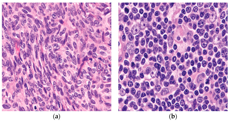
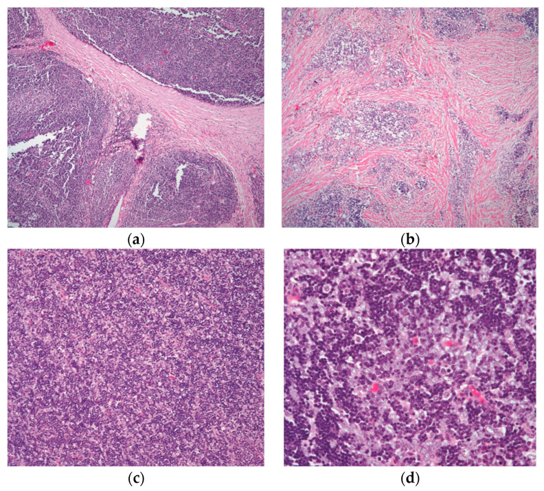
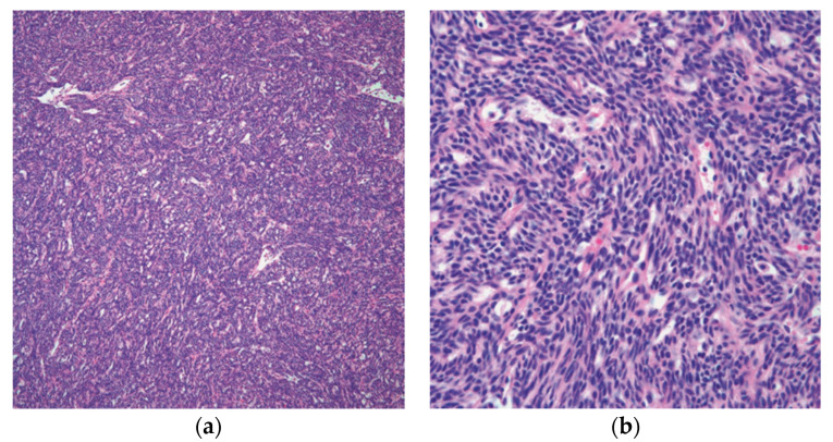
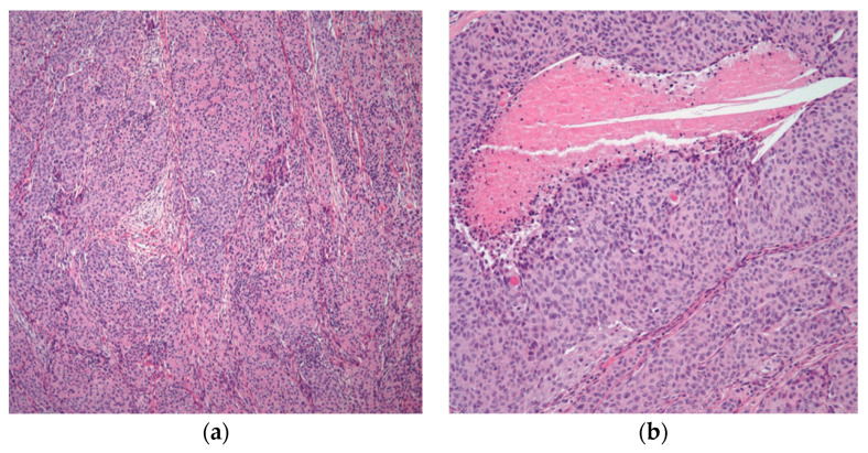
Thymomas are considered one of the most prevalent types of mediastinal epithelial tumors, which frequently develop in the anterior mediastinum. Due to their rarity, these tumors' nomenclature, classification, and staging are likely to be the subject of debate and argument for most expert pathologists. Furthermore, the significance of thymoma histologic classifications have been debated over the past twenty years. While certain advocates argue that staging at the time of diagnosis is more significant, others believe that histologic subtyping has a significant impact on how patients behave clinically. In this review, we will focus on some of the challenges that diagnostic surgical pathologists may experience while evaluating the histopathology of thymomas and staging these tumors. We will additionally glance over the clinical characteristics of these distinct tumors and the current management strategy.

Keywords: classification; mediastinum; staging; thymoma; thymus.


Conflict of interest statement

The author declares no conflict of interest.

Figures

Figure 1
Figure 1
The two epithelial components characteristically seen in thymoma: (a) the spindle/oval cells commonly seen in type A and type AB thymoma; (b) round/epithelioid epithelial cells found in type B thymoma. (a,b) (H&E, 40×).
Figure 2
Figure 2
The different growth patterns of conventional thymoma: (a) type B1 thymoma showing lobulation by thick fibrous bands; (b) thymoma in areas of fibrocollagenous stroma with no well-defined lobulation; (c) thymoma with an even distribution of lymphocytes and epithelial cells and a diffuse development pattern; and (d) higher magnification displaying the mixed population of lymphocytes and polygonal epithelial cells. (a,b) (H&E, 4×); (c) (H&E, 10×); (d) (H&E, 20×).
Figure 3
Figure 3
Spindle cell thymoma histologic feature: (a) low power of thymoma type A/spindle cell thymoma; (b) high power view showing the characteristic spindle cell cytology. (a) (H&E, 10×); (b) (H&E, 20×).
Figure 4
Figure 4
Atypical thymoma/WHO type B3: (a) atypical thymoma case showing nested pattern and rosette-like formation; (b) higher magnification showing atypia and focal comedo necrosis. (a) (H&E, 4×); (b) (H&E, 10×).
Figure 5
Figure 5
Micronodular thymoma with B-cell lymphoid hyperplasia: (a) islets of nodular epithelium embedded within a lymphocytic stroma; (b) many germinal centers are easily identifiable. (a) (H&E, 2×); (b) (H&E, 10×).

References

    1. Kalhor N., Moran C. Mediastinal Pathology. Springer; Berlin/Heidelberg, Germany: 2019.
    1. Suster S., Moran C.A. Thymoma classification: Current status and future trends. Am. J. Clin. Pathol. 2006;125:542–554. doi: 10.1309/CAV8RNU5TKNACKNC. - DOI - PubMed
    1. Anastasiadis K., Ratnatunga C. The Thymus Gland. Springer; Berlin/Heidelberg, Germany: 2007.
    1. Tsao M.-S., Nicholson A.G., Maleszewski J.J., Marx A., Travis W.D. Reprint of “Introduction to 2021 WHO Classification of Thoracic Tumors”. J. Thorac. Oncol. 2022;17:337–340. doi: 10.1016/j.jtho.2022.01.008. - DOI - PubMed
    1. Rich A.L. Epidemiology of thymoma. J. Thorac. Dis. 2020;12:7531. doi: 10.21037/jtd-2019-thym-02. - DOI - PMC - PubMed

LinkOut - more resources