. 2023 Aug 30;14(9):1733.
doi: 10.3390/genes14091733.

Mechanisms of Regulation of the CHRDL1 Gene by the TWIST2 and ADD1/SREBP1c Transcription Factors

Affiliations

Mechanisms of Regulation of the CHRDL1 Gene by the TWIST2 and ADD1/SREBP1c Transcription Factors

José J Casasnovas-Nieves et al. Genes (Basel). .

Abstract

Setleis syndrome (SS) is a rare focal facial dermal dysplasia caused by recessive mutations in the basic helix-loop-helix (bHLH) transcription factor, TWIST2. Expression microarray analysis showed that the chordin-like 1 (CHRDL1) gene is up-regulated in dermal fibroblasts from three SS patients with the Q119X TWIST2 mutation.

Methods: Putative TWIST binding sites were found in the upstream region of the CHRDL1 gene and examined by electrophoretic mobility shift (EMSA) and reporter gene assays.

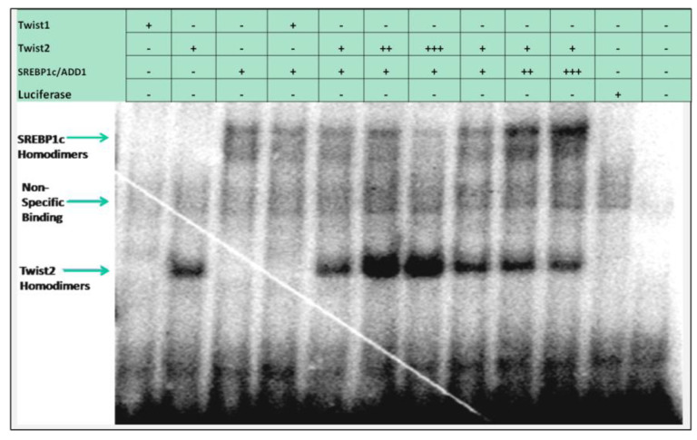
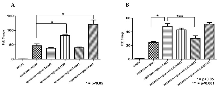
Results: EMSAs showed specific binding of TWIST1 and TWIST2 homodimers, as well as heterodimers with E12, to the more distal E-boxes. An adjoining E-box was bound by ADD1/SREBP1c. EMSA analysis suggested that TWIST2 and ADD1/SREBP1c could compete for binding. Luciferase (luc) reporter assays revealed that the CHRDL1 gene upstream region drives its expression and ADD1/SREBP1c increased it 2.6 times over basal levels. TWIST2, but not the TWIST2-Q119X mutant, blocked activation by ADD1/SREBP1c, but overexpression of TWIST2-Q119X increased luc gene expression. In addition, EMSA competition assays showed that TWIST2, but not TWIST1, competes with ADD1/SREBP1c for DNA binding to the same site.

Conclusions: Formation of an inactive complex between the TWIST2 Q119X and Q65X mutant proteins and ADD1/SREBP1c may prevent repressor binding and allow the binding of other regulators to activate CHRDL1 gene expression.

Keywords: ADD1; BMP signaling; CHRDL1; SREBP1c; Setleis syndrome; TWIST transcription factors; bHLH.


Conflict of interest statement

The authors declare no conflict of interest. The funders had no role in the design of the study; in the collection, analyses, or interpretation of data; in the writing of the manuscript; or in the decision to publish the results.

Figures

Figure 1
Figure 1
Schematic representation of the putative binding sites identified by the bioinformatic analysis using TESS. Five putative TWIST binding sites were identified in the 5′ upstream region, relative to the transcription start site of the CHRDL1 gene. In addition, putative binding sites for other proteins known to interact with TWIST2 were also predicted by TESS. The distance between sites is not in scale, but the diagram shows the relative positions of the putative binding sites. Based on this analysis we designed five probes, shown here as red bars, for carrying out DNA binding assays by EMSA. Probe #1 comprises nucleotides from −240 to −58, probe #2 comprises from −1044 to −917, probe #3 comprises from −1297 to −1148, probe #4 comprises from −2297 to −2198, and probe #5 comprises from −2697 to −2548 (relative to the transcription start site in human reference sequence NG_012816 REGION: 2242.127203).
Figure 2
Figure 2
Electrophoretic mobility shift assay for the most upstream region of the CHRDL1 gene tested with TWIST1 and TWIST2 proteins. Typical results of EMSA carried out with the in vitro synthesized TWIST proteins using probe #5. The table above the figure indicates with a plus (+) sign the proteins expressed in the TnT extracts and the unlabeled oligonucleotide competitors added to the DNA binding reactions. A minus (−) sign indicates the absence of an expressed protein or competitor oligonucleotide in the reaction. SP = specific oligonucleotide competitor; NS = non-specific oligonucleotide competitor. The (top image) represents a 15 h exposure, while the (bottom image) is from a 36 h exposure to X-ray film. Both TWIST1 and TWIST2 homodimers were able to bind this probe, but binding of the TWIST2 Q119X mutant protein could only be detected when overexposing the dried gel (bottom image). The binding is specific due to being competed out by an unlabeled probe but not by the unlabeled Sp1 element probe. Another shift is observed close to the wells’ origin but is considered non-specific binding as it is also observed in the luciferase lane (mock reaction).
Figure 2
Figure 2
Electrophoretic mobility shift assay for the most upstream region of the CHRDL1 gene tested with TWIST1 and TWIST2 proteins. Typical results of EMSA carried out with the in vitro synthesized TWIST proteins using probe #5. The table above the figure indicates with a plus (+) sign the proteins expressed in the TnT extracts and the unlabeled oligonucleotide competitors added to the DNA binding reactions. A minus (−) sign indicates the absence of an expressed protein or competitor oligonucleotide in the reaction. SP = specific oligonucleotide competitor; NS = non-specific oligonucleotide competitor. The (top image) represents a 15 h exposure, while the (bottom image) is from a 36 h exposure to X-ray film. Both TWIST1 and TWIST2 homodimers were able to bind this probe, but binding of the TWIST2 Q119X mutant protein could only be detected when overexposing the dried gel (bottom image). The binding is specific due to being competed out by an unlabeled probe but not by the unlabeled Sp1 element probe. Another shift is observed close to the wells’ origin but is considered non-specific binding as it is also observed in the luciferase lane (mock reaction).
Figure 3
Figure 3
Electrophoretic Mobility Shift Assay for the most upstream region of the CHRDL1 gene with TWIST, E12 and SREBP1c bHLH proteins. EMSA carried out with probe #5 to assess the SREBP1 putative binding site adjacent to the TWIST binding sites. The table above the gel image indicates the protein or oligonucleotide reagents added (plus (+) sign) or not included (minus (−) sign) in the DNA binding reactions. Since we observed binding of TWIST1 and TWIST2 homodimers to this probe, we assessed whether the SREBP1 putative binding site was bound by SREBP1c since this transcription factor is known to interact with TWIST2. A shift for SREBP1c was detected and confirmed by a supershift reaction accomplished using an antibody against the c-Myc tag of SREBP1c. In addition, we detected what could be intermediate complexes of SREBP1c /TWIST2, but their stoichiometry was not clear.
Figure 4
Figure 4
EMSA using the CH2700EA double-stranded oligonucleotide. For this probe, we detected binding of both TWIST2 and SREBP1c homodimers. The table above the gel image indicates the protein or oligonucleotide reagents added (plus (+) sign) or not included (minus (−) sign) in the DNA binding reactions. As seen before, no binding of Q119X homodimers was detected. Probe #5 was used as a positive control for the EMSA binding reactions.
Figure 5
Figure 5
EMSA assessing the E-box preference for SREBP1c and TWIST2 using the CH2700EA oligonucleotide as probe. The table above the gel image indicates the protein or oligonucleotide reagents added (plus (+) sign) or not included (minus (−) sign) in the DNA binding reactions. Both homodimers of TWIST2 and SREBP1c were able to bind the CH2700EA oligonucleotide. In addition, we confirmed that these proteins do not bind the −2611 site since the CH2600TWI oligonucleotide did not compete out the binding of both proteins. When the CH2700EMAW oligonucleotide was used as a competitor, the shifts for SREBP1c and TWIST2 were competed out. Interestingly, when the CH2700EWAM oligonucleotide was used, it competed out the binding of SREBP1c but only competed partially for the binding of TWIST2. While SREBP1c seems to be able to bind both E-boxes, TWIST2 may bind strongly the −2648 E-box since it was not competed out completely.
Figure 6
Figure 6
EMSA of the CH2700EA oligonucleotide with varying amounts of TWIST2 and SREBP1c. To evaluate whether TWIST2 and SREBP1c can bind simultaneously to the same region or if both proteins compete for binding, EMSAs were carried out varying the amount of these two bHLH factors. The table above the gel image indicates the protein or oligonucleotide reagents added (plus (+) sign) or not included (minus (−) sign) in the DNA binding reactions. When including increasing amounts of a given protein’s TnT reaction, two or three + signs were indicated in the table above the gel. In this EMSA, we first tested binding of both SREBP1c and TWIST2 homodimers individually. When we kept a constant amount of SREBP1c and increased the amount of TWIST2, the shift for SREBP1c was weakened. The opposite was also true; in the presence of increasing amounts of SREBP1c, the binding of TWIST2 decreased. These results suggest that both proteins compete for the same binding site. TWIST1 binding to this oligonucleotide was weakly detected.
Figure 7
Figure 7
EMSA of TWIST/E12 heterodimers for evaluation of binding to probe #5. In vitro coupled transcription/translation reactions were set up using a plasmid containing the cDNA for TWIST1, TWIST2 or Q119X, combined with another plasmid containing the E12 coding sequence. The proteins obtained from these reactions were used in EMSA binding reactions containing probe #5. In this EMSA, we observed both homodimeric and heterodimeric binding. We detected binding of TWIST/E12 heterodimers, TWIST2/E12 heterodimers, and TWIST2-Q119X/E12 heterodimers. These mobility shifts were confirmed to be specific since they were competed out by the CH2700EA double stranded oligonucleotide used as a specific competitor, but the SP1 oligonucleotide did not compete. Supershift reactions confirmed that the complexes detected were from the in vitro translated proteins.
Figure 8
Figure 8
The N-terminus of TWIST2 is involved in SREBP1c’s reduced DNA-binding activity. (A) Diagram of how proteins were synthesized in vitro through coupled transcription/translation for the binding reactions to be used in electrophoretic mobility shift assays (EMSA). (B) Western Blot of SDS-PAGE of TnT reactions of co-expressed TWIST proteins and SREBP1c, detected with an anti-Myc antibody. Luciferase was used as a positive control for protein synthesis and as a negative control (mock) for EMSA binding reactions. (C) EMSA analysis for determining the DNA binding ability of each protein to the DNA. Mock reaction represents the firefly luciferase negative control. Free probe represents unbound labeled DNA.
Figure 9
Figure 9
TWIST2 uses its second conserved sub-motif (SEEE) to regulate the DNA-binding ability of SREBP1c in vitro. EMSA analysis was carried out to determine the DNA-binding ability of each protein, as well as to detect how co-expression of TWIST2 or its mutant forms, alter the DNA binding activity of the SREBP1c transcription factor to the CHRDL1 gene probe #5 region.
Figure 10
Figure 10
Production of TWIST1 and deletion mutant TWIST1 proteins using TnT reactions and assessment of DNA binding of TWIST1 proteins and SREBP1c to the CHRDL1 gene probe #5 region. (A) To confirm protein expression and stability, duplicate in vitro TnT reactions were analyzed by SDS-PAGE and autoradiography of proteins labeled with 35S-Methionine. (B) EMSA analysis of TWIST1 and deletion mutants. Homodimers mixed reactions (reactions colored in red): Co-expressed reactions (reactions colored in blue). Addition of anti-Myc antibody to confirm the presence of the SREBP1c-DNA complexes. Firefly luciferase (lane 19) was used as a negative control for EMSA. Free probe (lane 20) represents labeled DNA not bound by protein.
Figure 11
Figure 11
Luciferase reporter gene assay of the -3KCHRDL1-pGL4 construct to assess the effect of different bHLH proteins on the CHRDL1 gene upstream region. Transient transfection of GM00637 SV-40 transformed human fibroblast cells was performed as described in the Section 2. (A) The upstream region of CHRDL1 (from −2751 to +85) was able to drive expression of the -3KCHRDL1-pGL4 reporter construct, indicative of promoter activity. TWIST1 and TWIST2 reduced the basal expression of the reporter gene, but this reduction was not significant. Both SREBP1c and TWIST2 Q119X were able to significantly increase luciferase activity, indicating that both had an activating effect. (B) Luciferase reporter gene assays to assess the effect of TWIST1, TWIST2 and Q119X on SREBP1c-mediated activation. TWIST2 but not TWIST1 was able to significantly block activation by SREBP1c, suggesting that this inhibition may be specific for TWIST2. The TWIST2 Q119X mutant was unable to block SREBP1c activation, as expected. Statistical analysis for experiments presented in A and B were performed using Student’s T-test (N = 3). Error bars represent the standard error of the mean (SEM).
Figure 12
Figure 12
The expression of the -3KCHRDL1-pGL4 reporter gene construct is significantly reduced with sodium butyrate (NaBt) treatment. Approximately 1 × 10⁶ COS7 cells were transiently transfected or co-transfected with the indicated expression vectors. Transfection media were removed after 4 h of incubation and replaced with media. Sodium butyrate (1 mM) was added directly to cells 24 h prior to cell lysis and incubated overnight. Luciferase activity was normalized relative to the basal promoter activity. Co-expression of the transcription factors increased the expression of the reporter gene. Addition of NaBt significantly reduced the expression of the reporter gene. Transfection experiments were performed in duplicate and repeated independently five times (N = 5). Statistical analysis was performed using one-way ANOVA followed by unpaired Student’s T-test. Error bars represent standard error of the mean (SEM). * = p < 0.05; **** = p < 0.0001.
Figure 13
Figure 13
Adaptation of the model reported by the Spicer group of the bi-functional role of TWIST2 in the regulation of the CHRDL1 gene. (A) As seen for thrombospondin [51] homodimers of TWIST2 might repress CHRDL1 gene expression while TWIST2/E12 heterodimers might activate expression. The expression state of the CHRDL1 gene at a given moment would be determined by the abundance of either homo- or heterodimers. (B) In Setleis syndrome patients harboring the Q119X, the mutant homodimers appear to be non-functional. That is not the case for the Q119X/E12 heterodimers, which are able to activate the CHRDL1 gene. Hence, activation of CHRDL1 might occur even in situations where the homodimer to heterodimer ratio is high. Two alternatives to this model are that: (1) Q119X homodimers bind weakly and could be easily displaced by SREBP1c leading to CHRDL1 activation, and (2) Q119X might sequester other HLH repressors in a similar manner to the Id proteins.

References

    1. Setleis H., Kramer B., Valcarcel M., Einhorn A.H. Congenital ectodermal dysplasia of the face. Pediatrics. 1963;32:540–548. doi: 10.1542/peds.32.4.540. - DOI - PubMed
    1. Clark R.D., Golabi M., Lacassie Y., Hall B., Seto S. Expanded phenotype and ethnicity in Setleis syndrome. Am. J. Med. Genet. 1989;34:354–357. doi: 10.1002/ajmg.1320340311. - DOI - PubMed
    1. Artlich A., Schwinger E., Meinecke P. Setleis (bitemporal ‘forceps marks’) syndrome in a German family: Evidence for autosomal dominant inheritance. Clin. Dysmorphol. 1992;1:157–160. doi: 10.1097/00019605-199207000-00006. - DOI - PubMed
    1. Matsumoto S., Kuno T., Hamasaki Y., Miyazaki S., Miyabara S., Narisawa Y. Setleis bitemporal “Forceps Marks” Syndrome and its pathogenesis: A case report. Acta Paediatr. Jpn. 1991;33:186–190. doi: 10.1111/j.1442-200X.1991.tb01541.x. - DOI - PubMed
    1. McGaughran J., Aftimos S. Setleis syndrome: Three new cases and a review of the literature. Am. J. Med. Genet. 2002;111:376–380. doi: 10.1002/ajmg.10632. - DOI - PubMed

LinkOut - more resources