Review
. 2023 Sep 6;24(18):13717.
doi: 10.3390/ijms241813717.

Analysis of the Structural Dynamics of Proteins in the Ligand-Unbound and -Bound States by Diffracted X-ray Tracking

Affiliations
Review

Analysis of the Structural Dynamics of Proteins in the Ligand-Unbound and -Bound States by Diffracted X-ray Tracking

Masayuki Oda. Int J Mol Sci. .

Abstract

Although many protein structures have been determined at atomic resolution, the majority of them are static and represent only the most stable or averaged structures in solution. When a protein binds to its ligand, it usually undergoes fluctuation and changes its conformation. One attractive method for obtaining an accurate view of proteins in solution, which is required for applications such as the rational design of proteins and structure-based drug design, is diffracted X-ray tracking (DXT). DXT can detect the protein structural dynamics on a timeline via gold nanocrystals attached to the protein. Here, the structure dynamics of single-chain Fv antibodies, helix bundle-forming de novo designed proteins, and DNA-binding proteins in both ligand-unbound and ligand-bound states were analyzed using the DXT method. The resultant mean square angular displacements (MSD) curves in both the tilting and twisting directions clearly demonstrated that structural fluctuations were suppressed upon ligand binding, and the binding energies determined using the angular diffusion coefficients from the MSD agreed well with the binding thermodynamics determined using isothermal titration calorimetry. In addition, the size of gold nanocrystals is discussed, which is one of the technical concerns of DXT.

Keywords: DNA-binding protein; antibody; binding energy; fluctuation; helix-bundle protein.


Conflict of interest statement

The author declares no conflict of interest.

Figures

Figure 1
Figure 1
Crystal structure of C6 scFv in complex with NP (PDB code; 6K4Z). The light and heavy chains of C6 scFv are indicated in cyan and green, respectively. The antigen, NP (magenta), and the side-chain of the 54th residue (Asn) of the heavy chain are indicated by the stick-model. The figure was drawn by PyMOL 2.5.2 (Schrodinger, LLC, New York, NY, USA).
Figure 2
Figure 2
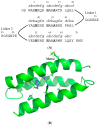
(A) Amino acid sequence of HA [30]. The His residues involved in metal ion binding and the Ala residues changed from hydrophobic amino acids of α3D are indicated in bold. The positions a—g are also indicated above the amino acids. (B) Structure model of HA based on NMR structure of α3D (PDB code; 2A3D). The side-chains of His and Ala in the hydrophobic core and that of Met64 are indicated by the stick-model. The figure was drawn by PyMOL 2.5.2 (Schrodinger, LLC, New York, NY, USA).
Figure 3
Figure 3
NMR structure of c-Myb R2R3 in complex with DNA (PDB code; 1MSE). The residue His135 is mutated to Met and its side-chain is indicated by stick-model. The figure was drawn by PyMOL 2.5.2 (Schrodinger, LLC, New York, NY, USA).
Figure 4
Figure 4
Histograms of the Δ absolute angular displacement of gold nanocrystal on R2R3 mutant C130I/H135M/M189A in θ direction within 100 ms. The histograms were compared in the presence of DNA at low (A) and high (B) immobilization densities. The figures were slightly modified from Figure 1A,B in [32].

Similar articles

Cited by

References

    1. Velankar S., Burley S.K., Kurisu G., Hoch J.C., Markley J.L. Structural Proteomics. Volume 2305. Humana; New York, NY, USA: 2021. The Protein Data Bank archive; pp. 3–21. Methods in Molecular Biology. - DOI - PubMed
    1. Kurisu G., Bekker G.J., Nakagawa A. History of Protein Data Bank Japan: Standing at the beginning of the age of structural genomics. Biophys. Rev. 2022;14:1233–1238. doi: 10.1007/s12551-022-01021-w. - DOI - PMC - PubMed
    1. Burley S.K., Berman H.M., Chiu W., Dai W., Flatt J.W., Hudson B.P., Kaelber J.T., Khare S.D., Kulczyk A.W., Lawson C.L., et al. Electron microscopy holdings of the Protein Data Bank: The impact of the resolution revolution, new validation tools, and implications for the future. Biophys. Rev. 2022;14:1281–1301. doi: 10.1007/s12551-022-01013-w. - DOI - PMC - PubMed
    1. Hoch J.C., Baskaran K., Burr H., Chin J., Eghbalnia H.R., Fujiwara T., Gryk M.R., Iwata T., Kojima C., Kurisu G., et al. Biological Magnetic Resonance Data Bank. Nucleic Acids Res. 2023;51:D368–D376. doi: 10.1093/nar/gkac1050. - DOI - PMC - PubMed
    1. Ishima R., Kurt Yilmaz N., Schiffer C.A. NMR and MD studies combined to elucidate inhibitor and water interactions of HIV-1 protease and their modulations with resistance mutations. J. Biomol. NMR. 2019;73:365–374. doi: 10.1007/s10858-019-00260-6. - DOI - PMC - PubMed

LinkOut - more resources