. 2023 Sep 6;24(18):13738.
doi: 10.3390/ijms241813738.

Crystal Structures of Bacterial Pectin Methylesterases Pme8A and PmeC2 from Rumen Butyrivibrio

Affiliations

Crystal Structures of Bacterial Pectin Methylesterases Pme8A and PmeC2 from Rumen Butyrivibrio

Vincenzo Carbone et al. Int J Mol Sci. .

Abstract

Pectin is a complex polysaccharide that forms a substantial proportion of the plant's middle lamella of forage ingested by grazing ruminants. Methanol in the rumen is derived mainly from methoxy groups released from pectin by the action of pectin methylesterase (PME) and is subsequently used by rumen methylotrophic methanogens that reduce methanol to produce methane (CH4). Members of the genus Butyrivibrio are key pectin-degrading rumen bacteria that contribute to methanol formation and have important roles in fibre breakdown, protein digestion, and the biohydrogenation of fatty acids. Therefore, methanol release from pectin degradation in the rumen is a potential target for CH4 mitigation technologies. Here, we present the crystal structures of PMEs belonging to the carbohydrate esterase family 8 (CE8) from Butyrivibrio proteoclasticus and Butyrivibrio fibrisolvens, determined to a resolution of 2.30 Å. These enzymes, like other PMEs, are right-handed β-helical proteins with a well-defined catalytic site and reaction mechanisms previously defined in insect, plant, and other bacterial pectin methylesterases. Potential substrate binding domains are also defined for the enzymes.

Keywords: Butyrivibrio; methane; methanol; pectin; pectin methylesterase; rumen.


Conflict of interest statement

The authors declare no conflict of interest.

Figures

Figure 1
Figure 1
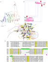
Crystal structure showing the predicted active and binding site domains of Pme8A (8TNE) from Butyrivibrio proteoclasticus B316T. (a) A ribbon representation of Pme8A monomer. The N- and C-terminal domains are indicated, including the presumptive active site of the enzyme. The unique elongated loops proximal to the active site (L1 and L2) are in purple. (b) A schematic diagram illustrating the protein’s topology in terms of how the β-strands (pink arrows) are arranged into β-sheets and α-helices (red cylinders). (c) A cartoon representation of the Pme8A monomer (in green) with L1 and L2 in purple and the superimposed bound hexasaccharide VI substrate of the pectin methylesterase 2NTP (light blue). Immediate active site and catalytic amino acids are shown in stick form for both enzymes and are labelled in accordance with the Pme8A structure. (d) Dali lite pair wise structural alignment of the larger crystal structure of Pme8A (monomer B) with the unique members of the larger PME enzyme superfamily (EC 3.1.1.11). Residues falling within 4.0–4.5 Å of the hexasaccharide VI substrate of 2NTP are highlighted in yellow for Pme8A as are the corresponding residues of the aligned enzymes. The unique elongated loops proximal to the presumptive active site (L1 and L2) are in purple. Residue numbering is in accordance with the Pme8A sequence.
Figure 2
Figure 2
Crystal structure showing the predicted active and binding site domains of PmeC2 (8TMS) from Butyrivibrio fibrisolvens D1T. (a) A ribbon representation of the PmeC2 monomer. The N- and C-terminal domains are indicated including the presumptive active site of the enzyme. The elongated loops proximal to the active site (L1 and L2) are in purple. (b) A schematic diagram illustrating the protein’s topology in terms of how the β-strands (pink arrows) are arranged into β-sheets and α-helices (red cylinders). (c) A cartoon representation of the PmeC2 monomer (in yellow) with the L1 and L2 in purple and the superimposed bound hexasaccharide VI substrate of the pectin methylesterase 2NTP in light blue. Immediate active site and catalytic amino acids are shown in stick form for both enzymes and labelled in accordance with the PmeC2 structure. (d) Dali lite pairwise structural alignment of the crystal structure of PmeC2 (monomer A) with the unique members of the larger PME enzyme superfamily (EC 3.1.1.11). Residues falling within 4.0–4.5 Å of the hexasaccharide VI substrate of 2NTP are highlighted in yellow for PmeC2 as are the corresponding residues of the aligned enzymes. The unique elongated loops proximal to the presumptive active site (L1 and L2) are in green. Residue numbering is in accordance with PmeC2 sequence.
Figure 3
Figure 3
Superimposed PmeC2 (in yellow, 8TMS) and Pme8A (in green, 8TNE) monomers. Secondary structural elements including loops (L1–L4) and N- and C-terminal α-helices are labelled.

References

    1. Morgavi D.P., Forano E., Martin C., Newbold C.J. Microbial ecosystem and methanogenesis in ruminants. Animal. 2010;4:1024–1036. doi: 10.1017/S1751731110000546. - DOI - PubMed
    1. Czerkawski J. Methane production in ruminants and its significance. World Rev. Nutr. Diet. 1969;11:240–282. - PubMed
    1. Janssen P.H. Influence of hydrogen on rumen methane formation and fermentation balances through microbial growth kinetics and fermentation thermodynamics. Anim. Feed Sci. Technol. 2010;160:1–22. doi: 10.1016/j.anifeedsci.2010.07.002. - DOI
    1. Kelly W.J., Leahy S.C., Kamke J., Soni P., Koike S., Mackie R., Seshadri R., Cook G.M., Morales S.E., Greening C., et al. Occurrence and expression of genes encoding methyl-compound production in rumen bacteria. Anim. Microbiome. 2019;1:1–13. doi: 10.1186/s42523-019-0016-0. - DOI - PMC - PubMed
    1. Kelly W.J., Mackie R.I., Attwood G.T., Janssen P.H., McAllister T.A., Leahy S.C. Hydrogen and formate production and utilisation in the rumen and the human colon. Anim. Microbiome. 2022;4:22. doi: 10.1186/s42523-022-00174-z. - DOI - PMC - PubMed