. 2023 Sep 12;24(18):13994.
doi: 10.3390/ijms241813994.

Modulation of LPS-Induced Neurodegeneration by Intestinal Helminth Infection in Ageing Mice

Affiliations

Modulation of LPS-Induced Neurodegeneration by Intestinal Helminth Infection in Ageing Mice

Natalia Jermakow et al. Int J Mol Sci. .

Abstract

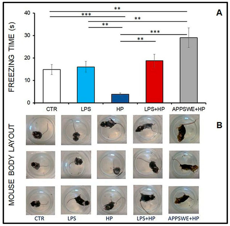
Parasitic helminths induce a transient, short-term inflammation at the beginning of infection, but in persistent infection may suppress the systemic immune response by enhancing the activity of regulatory M2 macrophages. The aim of the study was to determine how nematode infection affects age-related neuroinflammation, especially macrophages in the nervous tissue. Here, intraperitoneal LPS-induced systemic inflammation resulting in brain neurodegeneration was enhanced by prolonged Heligmosomoides polygyrus infection in C57BL/6 mice. The changes in the brain coincided with the increase in M1 macrophages, reduced survivin level, enhanced APP and GFAP expression, chitin-like chains deposition in the brain and deterioration behaviour manifestations. These changes were also observed in transgenic C57BL/6 mice predisposed to develop neurodegeneration typical for Alzheimer's disease in response to pathogenic stimuli. Interestingly, in mice infected with the nematode only, the greater M2 macrophage population resulted in better results in the forced swim test. Given the growing burden of neurodegenerative diseases, understanding such interactive associations can have significant implications for ageing health strategies and disease monitoring.

Keywords: ageing; helminths; immunomodulation; neurobehavioural manifestations; neurodegenerative diseases; neuroinflammation; persistent infection.


Conflict of interest statement

The authors declare that the research was conducted in the absence of any commercial or financial relationships that could be construed as a potential conflict of interest.

Figures

Figure 1
Figure 1
Changes in the percentage of neutrophils (A), monocytes (B) and eosinophils (C) in the blood smears of C57BL/6 mice, untreated, control (CTR), after injection with LPS (LPS), infection with Heligmosomoides polygyrus (HP) or injection with LPS and infection with H. polygyrus (LPS + HP). A statistically significant difference in the cell response: (A), ANOVA: between group, p < 0.002, the interaction between group and day, p < 0.0001; (B), ANOVA: between day, p < 0.000001; (C), ANOVA: between day, p < 0.00004, the interaction between group and day, p < 0.00002.
Figure 2
Figure 2
Changes in the cell population in the cerebrospinal fluid of C57BL/6 mice: untreated control (CTR), injected with LPS (LPS), infected with Heligmosomoides polygyrus (HP) or injected with LPS and infected with H. polygyrus (LPS + HP), and in APPSWE mice infected with H. polygyrus (APPSWE + HP). (A) Mean percentage of M1 and M2 macrophages. (B) Gating strategy for M1 and M2 analysis: all cells in the sample (P1), leukocytes negative for CD11b (P2), macrophages (P3) and microglia (P4) are distinguished based on the difference in CD11b and CD45 expression. Two populations of macrophages were identified in P3: M1 CD206+ CD80+ (Q2) and M2 CD206+ CD80− (Q4). (C) Representative dot plots. Statistical significance of differences between groups evaluated by Student t-test: * p < 0.05; ** p < 0.005; *** p < 0.00001. M1 ANOVA: between group, p < 0.0012; M2 ANOVA: between group, p < 0.00001.
Figure 3
Figure 3
The concentration of survivin in the brain homogenate in C57BL/6 mice: untreated, control (CTR), injected with LPS (LPS), infected with Heligmosomoides polygyrus (HP) or injected with LPS and infected with H. polygyrus (LPS + HP); APPSWE mice infected with H. polygyrus (APPSWE + HP). Samples were taken from 12-month-old mice infected with H. polygyrus at 4 months of age. Student t-test: ** p < 0.01; *** p < 0.001; ANOVA: between group, p < 0.000004.
Figure 4
Figure 4
Chitin-like polysaccharides in the brain of C57BL/6 mice: untreated, control (CTR), injected with LPS (LPS); infected with Heligmosomoides polygyrus (HP); injected with LPS and infected with H. polygyrus (LPS + HP); APPSWE mice infected with H. polygyrus (APPSWE + HP). (A) Brain slides with the positive reaction for extracellular chitin-like structure labelled by Calcofluor (DAPI channel at 60× magnification, with immersion). Mice non-infected with the nematode were negative. (B) The comparative pattern of FTIR spectra for chitin-like polysaccharides in brain homogenate samples. (C) Chitin-like polysaccharides with spectra typical for chitin. Samples were taken from 12-month-old mice infected with H. polygyrus at 4 months of age.
Figure 5
Figure 5
APP and GFAP co-detection in the brain of C56BL/6 mice: untreated, control mice (CTR); mice injected with LPS (LPS); mice infected with Heligmosomoides polygyrus (HP); mice injected with LPS and infected with H. polygyrus (LPS + HP) and APPSWE mice infected with H. polygyrus (APPSWE + HP). Atlas position of the tissue section in the brain (last column): thalamus (A,C), hippocampus (B,L), motor cortex (D,G,J,M), piriform cortex (E,H,K,N) and sensory cortex (F,I). Bright field, magnification 60×. The arrows in the LPS and HP groups indicate APP-positive neural cells. The arrows in the LPS + HP and APPSWE + HP groups indicate GFAP-positive astrocytes.
Figure 6
Figure 6
Western blotting for APP (A) and GFAP (B) in the brain of C57BL/6 mice: control mice (CTR), mice injected with LPS (LPS), mice infected with Heligmosomoides polygyrus (HP); mice injected with LPS and infected with H. polygyrus (LPS + HP) and APPSWE mice infected with H. polygyrus. The arrow indicates the α-tubulin position at 50 kDa. The rabbit anti-myeloid precursor protein (polyclonal antibodies AB5300 targeted against 27 amino acids (aa 99–126)) was used to detect the first amyloid β precursor; the typical set of bands corresponding to APP (95–100 kDa) in hippocampal (HIP) and cortex (COR) homogenates of 12-month-old mice. Additional smaller size bands in the hippocampus are also specific for APP, as they are not present in control (CTR) mice. They may be cytoplasmatic derivatives of APP with molecular weights between 24 and 28 kDa, indicating intensified protein cleavage. (C) Number of mice expressing long (100 kDa) and short (24–28 kDa) APP. The band protein was identified by Western blot and chemiluminescence. Not all mice in the group were positive in the cortex or hypothalamus for expression of short (24–28 kDa) APP chains. (D) Chemiluminescence intensity (%) of 100 kDa APP bands relative to 1 α-tubulin, a reference protein in brain tissue. The changes were not statistically significant. (E) Chemiluminescence intensity (%) of 55 kDa GFAP bands relative to 1 α-tubulin in brain tissue. The GFAP scores were statistically significant in the LPS + HP and APPSWE + HP groups in comparison to CTR, LPS and HP groups, in the cerebral cortex only. Student t-test, **** p < 0.0001; LPS + HP vs. CTR, LPS, HP; APPSWE + HP vs. CTR, LPS, HP.
Figure 7
Figure 7
Behavioural changes in the forced swim test of C57BL/6 mice: untreated, control (CTR); injected with LPS (LPS), infected with H. polygyrus (HP); injected with LPS and infected with H. polygyrus and in APPSWE mice infected with H. polygyrus (APPSWE + HP). (A) Average number of seconds (± standard error) of freezing. Student t-test: ** p < 0.01; *** p < 0.001; ANOVA: between group, p < 0.0000003. (B) Behavioural pattern of mice in forced swim test: In FST, mice placed in a container of water cannot escape. First, the animals tried to escape (CTR-active movement) or were passive and did not swim, but floated in the water and tried to keep their nose above the water (LPS + HP, APPSWE + HP). The swimming positions of mice and mouse body layouts from the LPS and HP groups changed to active at different intervals.

Similar articles

References

    1. Gupta P.P., Pandey R.D., Jha D., Shrivastav V., Kumar S. Role of traditional nonsteroidal anti-inflammatory drugs in Alzheimer’s disease: A meta-analysis of randomized clinical trials. Am. J. Alzheimer’s Dis. Other Demen. 2015;30:178–182. doi: 10.1177/1533317514542644. - DOI - PMC - PubMed
    1. Battaglia S., Di Fazio C., Vicario C.M., Avenanti A. Neuropharmacological Modulation of N-methyl-D-aspartate, Noradrenaline and Endocannabinoid Receptors in Fear Extinction Learning: Synaptic Transmission and Plasticity. Int. J. Mol. Sci. 2023;24:5926. doi: 10.3390/ijms24065926. - DOI - PMC - PubMed
    1. Batista C., Gomes G.F., Candelario-Jalil E., Fiebich B.L., de Oliveira A. Lipopolysaccharide-induced neuroinflammation as a bridge to understand neurodegeneration. Int. J. Mol. Sci. 2019;20:2293. doi: 10.3390/ijms20092293. - DOI - PMC - PubMed
    1. Catorce M.N., Gevorkian G. LPS-induced murine neuroinflammation model: Main features and suitability for pre-clinical assessment of nutraceuticals. Curr. Neuropharmacol. 2016;14:155–164. doi: 10.2174/1570159X14666151204122017. - DOI - PMC - PubMed
    1. Heppner F.L., Ransohoff R.M., Becher B. Immune attack: The role of inflammation in Alzheimer disease. Nat. Rev. Neurosci. 2015;16:358–372. doi: 10.1038/nrn3880. - DOI - PubMed

Substances

Supplementary concepts