. 2023 Sep 12;24(18):14004.
doi: 10.3390/ijms241814004.

Reduced Sphingosine in Cystic Fibrosis Increases Susceptibility to Mycobacterium abscessus Infections

Affiliations

Reduced Sphingosine in Cystic Fibrosis Increases Susceptibility to Mycobacterium abscessus Infections

Fabian Schnitker et al. Int J Mol Sci. .

Abstract

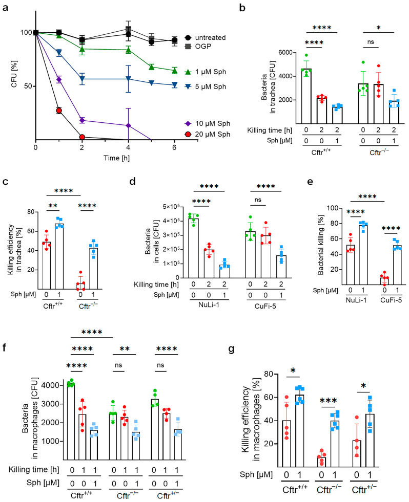
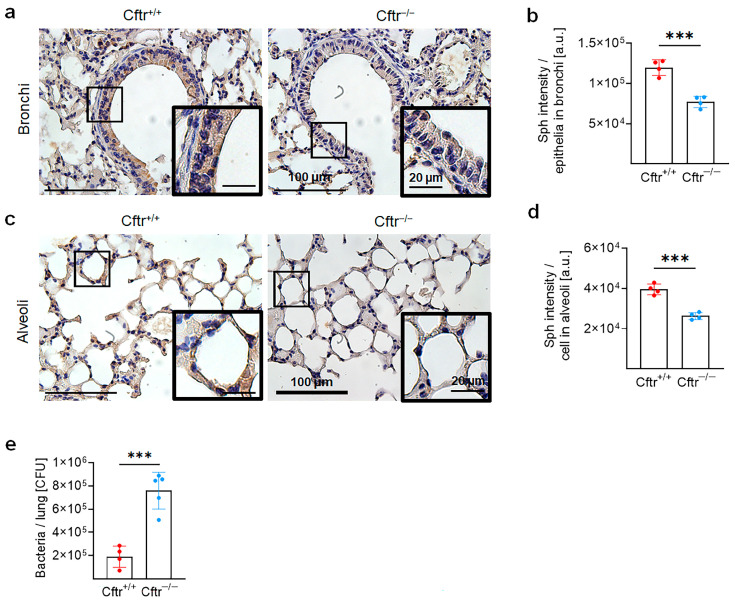
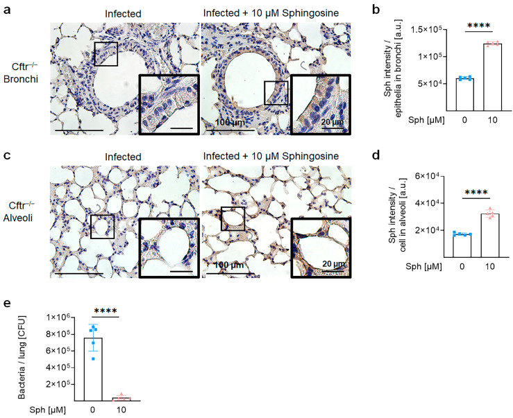
Cystic fibrosis (CF) is an autosomal recessive disorder caused by the deficiency of the cystic fibrosis transmembrane conductance regulator (CFTR) and often leads to pulmonary infections caused by various pathogens, including Staphylococcus aureus, Pseudomonas aeruginosa, and nontuberculous mycobacteria, particularly Mycobacterium abscessus. Unfortunately, M. abscessus infections are increasing in prevalence and are associated with the rapid deterioration of CF patients. The treatment options for M. abscessus infections are limited, requiring the urgent need to comprehend infectious pathogenesis and develop new therapeutic interventions targeting affected CF patients. Here, we show that the deficiency of CFTR reduces sphingosine levels in bronchial and alveolar epithelial cells and macrophages from CF mice and humans. Decreased sphingosine contributes to the susceptibility of CF tissues to M. abscessus infection, resulting in a higher incidence of infections in CF mice. Notably, treatment of M. abscessus with sphingosine demonstrated potent bactericidal activity against the pathogen. Most importantly, restoration of sphingosine levels in CF cells, whether human or mouse, and in the lungs of CF mice, provided protection against M. abscessus infections. Our findings demonstrate that pulmonary sphingosine levels are important in controlling M. abscessus infection. These results offer a promising therapeutic avenue for CF patients with pulmonary M. abscessus infections.

Keywords: Mycobacterium abscessus; cystic fibrosis; sphingolipids; sphingosine.


Conflict of interest statement

The authors declare no conflict of interest.

Figures

Figure 1
Figure 1
Cftr−deficient epithelial cells and macrophages are more susceptible to M. abscessus ssp. abscessus infection. (a,b) Tracheae from wildtype and Cftr—deficient mice were isolated and opened, and the epithelial surface was infected with 104 CFU of M. abscessus ssp. abscessus. After 2 h infection, the tracheae were washed, and the remaining bacteria were quantified through colony—forming unit assay (before killing). To allow killing of the bacteria, the infected and washed tracheae were further incubated in medium for 2 h and bacteria were determined through colony—forming unit assay (after killing). (b) The bacterial killing efficiency was calculated as described in the methods. (c,d) NuLi-1 and CuFi-5 cells were infected with 106 bacteria (MOI 10) for 2 h, washed, and CFU were determined (before killing). Infected and washed cells were further incubated in fresh medium for 2 h, and the bacterial number was determined as after killing. (d) The bacterial killing efficiency was then calculated. (e,f) Macrophages were derived from the bone marrow of wildtype, Cftr+/− or Cftr−/− mice and infected with 104 M. abscessus ssp. abscessus. After 2 h of infection, the cells were washed and the bacteria were quantified (before killing). The washed and infected cells were further incubated for 1 h. The bacterial number was then quantified as shown after killing (e). The killing efficiency of bacteria was then calculated (f). Shown are the mean ± SD of 4–5 (a,b), 5 (c,d), 4–5 (e,f) independent experiments; * p < 0.05, ** p < 0.01, *** p < 0.001, **** p < 0.0001, ns, not significant, t-test.
Figure 2
Figure 2
Sphingosine level is reduced in Cftr-deficient cells. Representative images of sphingosine staining and the intensity quantification by ImageJ Version 1.53t. (ac) NuLi-1 and CuFi-5 cells were either left uninfected (a) or infected with M. abscessus ssp. abscessus for 2 h, washed and further incubated for 2 h (b). (df) Bone-marrow-derived macrophages were isolated from Cftr+/+ or Cftr−/−mice, either left uninfected (d) or infected with M. abscessus ssp. abscessus for 2 h, washed and further incubated for 1 h (e). (c,f) Staining intensity in cells was determined by 10–20 cells per image. Three images from each experiment were quantified. The images were taken using light microscopy and analyzed with ImageJ Version 1.53t. Shown are the mean ± SD of 3 independent experiments; **** p < 0.0001, ns, not significant, one-way ANOVA followed by the Tukey test.
Figure 3
Figure 3
Sphingosine kills M. abscessus ssp. abscessus in vitro and intracellularly. (a) M. abscessus ssp. abscessus were resuspended in HEPES-buffered RPMI-1640 at a concentration of 106/mL with OGP (octyl-glucopyranoside, solvent of sphingosine) or sphingosine in different concentrations for the indicated time. The bacterial number was quantified from 5–9 independent experiments. (b,c) Tracheae isolated from wildtype or Cftr-deficient mice, (d,e) NuLi-1 and CuFi-5 cells or (f,g) bone-marrow-derived macrophages isolated from wildtype, Cftr+/−, or Cftr−/− were infected with M. abscessus ssp. abscessus for 2 h, washed, and CFU were determined and shown as killing for 0 h. The washed and infected cells were then incubated with fresh medium with or without 1 μM sphingosine for 2 h (epithelial cells) or 1 h (macrophages). The killing of bacteria was determined through a colony-forming unit assay. The panels (b,d,e) show the absolute CFU count, and the panels (c,e,g) show the killing efficiency calculated as described in the Section 4. Shown are the mean ± SD of 4–6 independent experiments, * p < 0.05, ** p < 0.01, *** p < 0.001, **** p < 0.0001, ns, not significant, 2-way ANOVA followed by the Tukey test.
Figure 4
Figure 4
Cftr-deficient mice are susceptible to M. abscessus ssp. abscessus infection and are associated with downregulated sphingosine levels in the lungs. (ad) The lungs of wildtype (Cftr+/+) or Cftr-deficient (Cftr−/−) mice were harvested, embedded in paraffin, and subjected to immunohistochemistry using anti-sphingosine antibodies. Panels (b,d) represent the quantification of staining in the bronchi (b) and alveoli (d). Staining intensity in the bronchi was quantified in the entire epithelial layer. Staining intensity in the alveoli was determined in five alveoli per image. From each mouse, 4–7 images were quantified and the average images were taken, n = 4. The images were taken using light microscopy and analyzed with ImageJ Version 1.53t. Brown staining indicates positive staining. Shown are mean ± SD, *** p < 0.001, t-test. Representative images from 4 mice are shown. (e) Wildtype or Cftr-deficient mice were intranasally infected with 106 CFU M. abscessus ssp. abscessus for 6 h and then sacrificed. The lungs were isolated and homogenized and the bacterial load was determined through colony-forming unit assays. Shown are the mean ± SD from n = 4–5 independent experiments. Statistical significance was determined through the t-test, *** p < 0.001.
Figure 5
Figure 5
Sphingosine treatment improves killing of M. abscessus ssp. abscessus in Cftr-deficient mice. Cftr-deficient mice were infected with M. abscessus ssp. abscessus (ad) for 60 min and then inhaled with either NaCl (square) or sphingosine (triangle). The lungs were isolated 6 h after infection. Panels (a,c) display representative immunohistochemistry staining of sphingosine from the bronchi (a) and alveoli (c). Images represent 5 independent experiments and were quantified using Image, J (b,d). (e) Mice were infected with M. abscessus ssp. abscessus for 60 min and then inhaled with 10 μM sphingosine or left untreated; the lungs were removed after 6 h and homogenized. Bacterial numbers in the lungs were determined through a colony-forming unit assay, n = 5, one-way ANOVA, and the Dunnett test. Shown are the mean ± SD of 5 independent experiments; **** p < 0.0001, t-test.
Figure 6
Figure 6
Sphingosine ameliorates M. abscessus ssp. abscessus infection in human ex vivo bronchi and lungs. (ad) Freshly isolated bronchi from donors and patients were either left uninfected (dot) or infected with M. abscessus ssp. abscessus for 1 h, washed, and then treated with 1 μM sphingosine (triangle) for 2 h or left untreated (square) and also further incubated for 2 h. (a) Bronchi were then washed and homogenized, and intracellular bacteria from patients or healthy donor bronchus were quantified through colony-forming unit assay. (b) Sphingosine in uninfected or infected donor bronchi was visualized using anti-sphingosine antibodies and immunohistochemistry staining. Representative results from five experiments are shown. (c) Staining intensities in 3–4 sections from 5 bronchi were quantified using ImageJ Version 1.53t. (d) Uninfected and infected donor bronchi were embedded in paraffin and sectioned, and the sections were stained with H&E. Arrows show the epithelial layer. Shown are representatives from five independent experiments. (e) Single cells were prepared from freshly isolated patient lung parenchyma and infected with M. abscessus ssp. abscessus for 1 h. Cells were then treated with sphingosine for 2 h or left untreated. Finally, the cells were extensively washed and intracellular bacteria were determined through colony-forming assay. The results are from every n = 9 sample. Shown are the mean ± SD; *** p < 0.001, **** p < 0.0001 2-way ANOVA, followed by the Tukey test or, if appropriate, using the t-test.

Similar articles

Cited by

References

    1. Elborn J.S. Cystic fibrosis. Lancet. 2016;388:2519–2531. doi: 10.1016/S0140-6736(16)00576-6. - DOI - PubMed
    1. Gadsby D.C., Vergani P., Csanady L. The ABC protein turned chloride channel whose failure causes cystic fibrosis. Nature. 2006;440:477–483. doi: 10.1038/nature04712. - DOI - PMC - PubMed
    1. Lopes-Pacheco M. CFTR Modulators: The Changing Face of Cystic Fibrosis in the Era of Precision Medicine. Front. Pharmacol. 2019;10:1662. doi: 10.3389/fphar.2019.01662. - DOI - PMC - PubMed
    1. Riordan J.R. CFTR function and prospects for therapy. Annu. Rev. Biochem. 2008;77:701–726. doi: 10.1146/annurev.biochem.75.103004.142532. - DOI - PubMed
    1. Riordan J.R. Assembly of functional CFTR chloride channels. Annu. Rev. Physiol. 2005;67:701–718. doi: 10.1146/annurev.physiol.67.032003.154107. - DOI - PubMed

MeSH terms

Substances

LinkOut - more resources