. 2023 Sep 14;24(18):14104.
doi: 10.3390/ijms241814104.

Catecholamines Promote Ovarian Cancer Progression through Secretion of CXC-Chemokines

Affiliations

Catecholamines Promote Ovarian Cancer Progression through Secretion of CXC-Chemokines

Hyun Jung Kim et al. Int J Mol Sci. .

Abstract

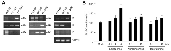
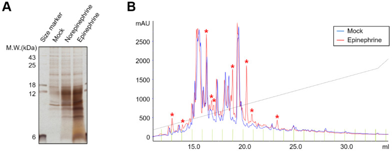
Considerable evidence has accumulated in the last decade supporting the notion that chronic stress is closely related to the growth, metastasis, and angiogenesis of ovarian cancer. In this study, we analyzed the conditioned media in SKOV3 ovarian cancer cell lines treated with catecholamines to identify secreted proteins responding to chronic stress. Here, we observed that epinephrine and norepinephrine enhanced the secretion and mRNA expression of CXC-chemokines (CXCL1, 2, 3, and 8). Neutralizing antibodies to CXCL8 and CXCL8 receptor (CXCR2) inhibitors significantly reduced catecholamine-mediated invasion of SKOV3 cells. Finally, we found that the concentration of CXCL1 and CXCL8 in the plasma of ovarian cancer patients increased with stage progression. Taken together, these findings suggest that stress-related catecholamines may influence ovarian cancer progression through the secretion of CXC-chemokines.

Keywords: CXCL1; CXCL8; catecholamine; invasion; ovarian cancer.


Conflict of interest statement

The authors declare no conflict of interest. The funders have no role in the design of the study; in the collection, analysis, or interpretation of data; in the writing of the manuscript; or in the decision to publish the results.

Figures

Figure 1
Figure 1
Catecholamines induce invasion of ovarian cancer cells. (A) mRNA level of adrenergic receptors in ovarian cancer cells. Total RNA isolated from HIO-80, SKOV3, and OVCAR3 cells was analyzed by RT-PCR using human adrenergic receptors and GAPDH-specific primers. (B) 5 × 10⁴ SKOV3 cells were seeded in the upper chambers, with the lower chambers containing 0.1, 1, or 10 μM epinephrine, norepinephrine, or isoproterenol. After culturing for 24 h, the infiltrated cells were fixed, stained, and observed using an optical microscopy. Results are representative of two independent experiments.
Figure 2
Figure 2
Catecholamines activate protein secretion in SKOV3 cells. (A) PBS-, 10 μM norepinephrine-, or 10 μM epinephrine-treated SKOV3 culture supernatants were collected and proteins were separated by SDS-PAGE and visualized by silver staining. (B) PBS- or 10 μM epinephrine-treated SKOV3 culture supernatants were collected and performed using an Ultimate HPLC system. Total ion chromatograms of control (Mock, blue) and 10 μM epinephrine (Epi, red) treated culture medium. Peaks specifically increased by epinephrine-treatment are marked with asterisks.
Figure 3
Figure 3
Epinephrine induces CXC-chemokines in SKOV3 cells. (A) Levels of cytokines and chemokines in the SKOV3 culture media were determined with cytokine arrays after the cells were treated with vehicle control (top panel) and 10 μM of epinephrine (bottom panel) at 24 h. (B) Enlarged images of negative control, positive control, and significantly increased cytokines from (A).
Figure 4
Figure 4
Catecholamines induce secretion of CXCL1, CXCL2, CXCL3, and CXCL8 in ovarian cancer cells. 1 × 10⁶ SKOV3 (A,C) or OVCAR3 (B,D) cells in 6-well plate were treated with 0.1, 1, or 10 μM epinephrine, norepinephrine, or isoproterenol. After 24 h, the concentrations of CXCL1 (A,B) and CXCL8 (C,D) in the culture medium were measured using ELISA. (E,F) Time-dependent secretion of CXCL1 or CXCL8 in SKOV3 cells. After 0, 3, 6, 12, 24, 48 h in the presence or absence of 10 μM epinephrine in SKOV3 cells, conditioned medium was obtained and the concentrations of CXCL1 (E) and CXCL8 (F) were measured by ELISA. (G) Levels of CXCL2 and CXCL3 were determined by Western blot analysis using anti-CXCL2 or CXCL3 antibodies. The time-dependent expression changes of the target gene are represented as fold change relative to the initial time (0 h). (H) The time-dependent expression changes of the target gene are represented as fold change relative to the initial time (0 h) and were normalized to GAPDH. (H) Epinephrine induces the mRNA expression of CXCL1, CXCL2, CXCL3, and CXCL8 in SKOV3 cells. 1 × 10⁶ SKOV3 cells in 6-well plate were treated with 10 μM epinephrine and cells were harvested after 0, 6, 12, 24, and 48 h. Total RNA was isolated and RT-PCR analysis was carried out for CXCL1, CXCL2, CXCL3, and CXCL8. GAPDH is shown as a control for RNA loading. Results are representative of two independent experiments.
Figure 5
Figure 5
CXCL8 secreted by SKOV3 cells induces SKOV3 cell invasion. (A) mRNA levels of chemokines and chemokine receptors in ovarian cancer cells. Total RNA isolated from HIO-80, SKOV3, and OVCAR3 cells was analyzed by RT-PCR using chemokine (CXCL1, CXCL2, CXCL3, and CXCL8)- and chemokine receptor (CXCR1, CXCR2, and ACKR1)-specific primers. (B) 5 × 10⁴ SKOV3 cells were seeded into a matrigel-coated upper chamber with a lower chamber containing 10 μM epinephrine in the presence or absence of MAB208 (CXCL8 neutralizing antibody), SB225002, or SB265610. After culturing for 24 h, infiltrated cells were fixed, stained, and observed using an optical microscopy. Results are representative of two independent experiments.
Figure 6
Figure 6
Elevated levels of CXCL1 and CXCL8 in plasma samples from ovarian cancer patients. Scatter plots show the concentrations of CXCL1 (A) and CXCL8 (B) in plasma samples from ovarian cancer patients at each stage of cancer. Analysis of CXCL1 and CXCL8 in plasma samples from ovarian cancer patients and normal blood donors were measured using ELISA. The horizontal bars represent the median and whiskers indicate the interquartile range. The healthy patients were used as the control group for statistical analysis. The p-value was calculated using one-way ANOVA, **** p < 0.0001, ** p < 0.01 and * p < 0.05.

Similar articles

Cited by

References

    1. Segerstrom S.C., Miller G.E. Psychological stress and the human immune system: A meta-analytic study of 30 years of inquiry. Psychol. Bull. 2004;130:601–630. doi: 10.1037/0033-2909.130.4.601. - DOI - PMC - PubMed
    1. Degroote C., Schwaninger A., Heimgartner N., Hedinger P., Ehlert U., Wirtz P.H. Acute Stress Improves Concentration Performance. Exp. Psychol. 2020;67:88–98. doi: 10.1027/1618-3169/a000481. - DOI - PubMed
    1. Liu Y., Tian S., Ning B., Huang T., Li Y., Wei Y. Stress and cancer: The mechanisms of immune dysregulation and management. Front. Immunol. 2022;13:1032294. doi: 10.3389/fimmu.2022.1032294. - DOI - PMC - PubMed
    1. Mariotti A. The effects of chronic stress on health: New insights into the molecular mechanisms of brain-body communication. Future Sci. OA. 2015;1:FSO23. doi: 10.4155/fso.15.21. - DOI - PMC - PubMed
    1. Yan J., Chen Y., Luo M., Hu X., Li H., Liu Q., Zou Z. Chronic stress in solid tumor development: From mechanisms to interventions. J. Biomed. Sci. 2023;30:8. doi: 10.1186/s12929-023-00903-9. - DOI - PMC - PubMed

LinkOut - more resources