. 2023 Sep 15;24(18):14150.
doi: 10.3390/ijms241814150.

The Biocompatibility Analysis of Artificial Mucin-Like Glycopolymers

Affiliations

The Biocompatibility Analysis of Artificial Mucin-Like Glycopolymers

P Trosan et al. Int J Mol Sci. .

Abstract

The ocular surface is covered by a tear film consisting of an aqueous/mucin phase and a superficial lipid layer. Mucins, highly O-glycosylated proteins, are responsible for lubrication and ocular surface protection. Due to contact lens wear or eye disorders, lubrication of the ocular surface can be affected. Artificial glycopolymers which mimic natural mucins could be efficient in ophthalmic therapy. Various neutral, positively, and negatively charged mucin-mimicking glycopolymers were synthesized (n = 11), cultured in different concentrations (1%, 0.1%, and 0.01% w/v) with human corneal epithelial cells (HCE), and analyzed by various cytotoxicity/viability, morphology, and immunohistochemistry (IHC) assays. Six of the eleven glycopolymers were selected for further analysis after cytotoxicity/viability assays. We showed that the six selected glycopolymers had no cytotoxic effect on HCE cells in the 0.01% w/v concentration. They did not negatively affect cell viability and displayed both morphology and characteristic markers as untreated control cells. These polymers could be used in the future as mucin-mimicking semi-synthetic materials for lubrication and protection of the ocular surface.

Keywords: cornea; epithelial corneal cells; glycopolymers; mucin.


Conflict of interest statement

The authors declare no conflict of interest.

Figures

Figure 1
Figure 1
Chemical structures of neutrally and negatively charged glycopolymers.
Figure 2
Figure 2
Chemical structures of cationic glycopolymers.
Figure 3
Figure 3
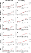
The cytotoxicity (A,C,E,G,I,K,M) and viability (B,D,F,H,J,L,N) assays of cultured HCE itself (control), with different glycopolymers (in 1, 0.1, and 0.01% w/v concentration) and PEI (MW 750k). The intensity of luminescence (viability; y axis) and fluorescence (cytotoxicity; y-axis) was measured at various time points (0–48 h; x-axis) by Microplate Reader. The data were normalized to control at time point 0. Interleaved symbols were used for every time point in the graphs.
Figure 4
Figure 4
Morphology of cultured HCE itself (control; (A)), with PGalNAcMAm (BD), PLacMAm-2 (EG), Lac-PEI-1.8k (HJ), Lac-PEI-10k (KM), S-Lac-1 (NP), S-Lac-2 (QS) and PEI (MW 750k; (TV)) in 1, 0.1 and 0.01% w/v concentration after 48 h. Light microscopy, scale bars represent 100 μm.
Figure 5
Figure 5
Comparison of the metabolic activity of the cultured HCE itself (control), with PGalNAcMAm (A), PLacMAm-2 (B), Lac-PEI-1.8k (C), Lac-PEI-10k (D), S-Lac-1 (E), S-Lac-2 (F) and PEI (MW 750k; (G)) in 1, 0.1 and 0.01% w/v concentration after 48 h. WST-8 reagent was added to the cell cultures for 4 h to form formazan. The absorbance was measured at a wave-length of 450 nm. * p < 0.05, *** p ≤ 0.001.
Figure 6
Figure 6
Comparison of the percentage of viable cultured HCE itself (control) with different glycopolymers (0.01% w/v concentration) and PEI (MW 750k). A Live-Dead assay was applied after 48 h. Live cells were stained with Calcein (green) and dead cells with Ethidium homodimer-1 (red). The scale bar presented in each image is 50 µm.
Figure 7
Figure 7
Immunostaining of proliferation marker Ki-67 (red) in cultured HCE itself (Control) with different glycopolymers (in 1, 0.1, and 0.01% w/v concentration) and PEI (MW 750k). Cell nuclei were counterstained with DAPI (blue). The scale bar represents 50 μm.
Figure 8
Figure 8
Immunostaining of markers of HCE (Pax6 (A); ABCG2 (B)) in cultured HCE itself (control) with different glycopolymers (in 1, 0.1, and 0.01% w/v concentration) and PEI (MW 750k). Cell nuclei were counterstained with DAPI (blue). The scale bar represents 50 μm.

References

    1. Tan D.T., Pullum K.W., Buckley R.J. Medical applications of scleral contact lenses: 1. A retrospective analysis of 343 cases. Cornea. 1995;14:121–129. doi: 10.1097/00003226-199503000-00002. - DOI - PubMed
    1. Tan D.T., Pullum K.W., Buckley R.J. Medical applications of scleral contact lenses: 2. Gas-permeable scleral contact lenses. Cornea. 1995;14:130–137. doi: 10.1097/00003226-199503000-00002. - DOI - PubMed
    1. Pullum K.W., Whiting M.A., Buckley R.J. Scleral contact lenses: The expanding role. Cornea. 2005;24:269–277. doi: 10.1097/01.ico.0000148311.94180.6b. - DOI - PubMed
    1. Foss A.J., Trodd T.C., Dart J.K. Current indications for scleral contact lenses. CLAO J. 1994;20:115–118. - PubMed
    1. Lim P., Ridges R., Jacobs D.S., Rosenthal P. Treatment of persistent corneal epithelial defect with overnight wear of a prosthetic device for the ocular surface. Am. J. Ophthalmol. 2013;156:1095–1101. doi: 10.1016/j.ajo.2013.06.006. - DOI - PubMed

LinkOut - more resources