

Structural and Thermomagnetic Properties of Gallium Nanoferrites and Their Influence on Cells In Vitro
- PMID: 37762487
- PMCID: PMC10532423
- DOI: 10.3390/ijms241814184
Structural and Thermomagnetic Properties of Gallium Nanoferrites and Their Influence on Cells In Vitro
Abstract
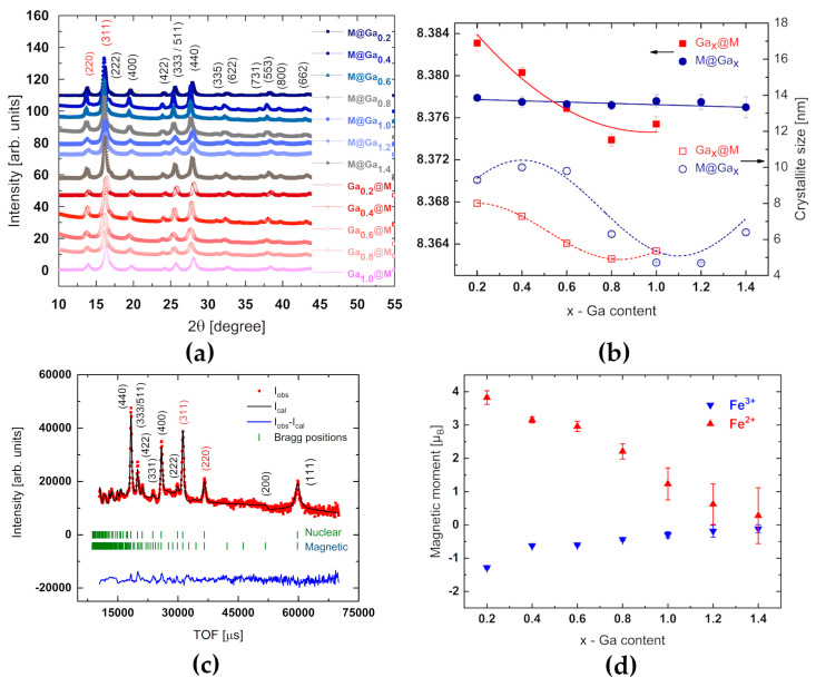
Magnetite and gallium substituted cuboferrites with a composition of GaxFe3-xO4 (0 ≤ x ≤ 1.4) were fabricated by thermal decomposition from acetylacetonate salts. The effect of Ga3+ cation substitution on the structural and thermomagnetic behavior of 4-12 nm sized core-shell particles was explored by X-ray and neutron diffraction, small angle neutron scattering, transmission electron microscopy, Mössbauer spectroscopy, and calorimetric measurements. Superparamagnetic (SPM) behavior and thermal capacity against increasing gallium concentration in nanoferrites were revealed. The highest heat capacity typical for Fe3O4@Ga0.6Fe2.4O4 and Ga0.6Fe2.4O4@Fe3O4 is accompanied by a slight stimulation of fibroblast culture growth and inhibition of HeLa cell growth. The observed effect is concentration dependent in the range of 0.01-0.1 mg/mL and particles of Ga0.6Fe2.4O4@Fe3O4 design have a greater effect on cells. Observed magnetic heat properties, as well as interactions with tumor and healthy cells, provide a basis for further biomedical research to use the proposed nanoparticle systems in cancer thermotherapy (magnetic hyperthermia).
Keywords: Mössbauer spectroscopy; X-ray and neutron diffraction; calorimetric measurements; in vitro cell culture; small angle neutron scattering; superparamagnetic nanoparticles.
Conflict of interest statement
The authors declare no conflict of interest.
Figures


Similar articles
-
Shell-mediated control of surface chemistry of highly stoichiometric magnetite nanoparticles.Nanoscale. 2020 Jul 2;12(25):13626-13636. doi: 10.1039/d0nr02069a. Nanoscale. 2020. PMID: 32558841
-
Insight into synthesis and characterisation of Ga0.9Fe2.1O4 superparamagnetic NPs for biomedical applications.Sci Rep. 2023 Oct 24;13(1):18175. doi: 10.1038/s41598-023-45285-y. Sci Rep. 2023. PMID: 37875541 Free PMC article.
-
Spin canting across core/shell Fe3O4/MnxFe3-xO4 nanoparticles.Sci Rep. 2018 Feb 21;8(1):3425. doi: 10.1038/s41598-018-21626-0. Sci Rep. 2018. PMID: 29467424 Free PMC article.
-
Two-step single-reactor synthesis of oleic acid- or undecylenic acid-stabilized magnetic nanoparticles by thermal decomposition.Beilstein J Nanotechnol. 2023 Jan 3;14:11-22. doi: 10.3762/bjnano.14.2. eCollection 2023. Beilstein J Nanotechnol. 2023. PMID: 36703905 Free PMC article.
-
Fabrication, optimization, and characterization of ultra-small superparamagnetic Fe3O4 and biocompatible Fe3O4@ZnS core/shell magnetic nanoparticles: Ready for biomedicine applications.Mater Sci Eng C Mater Biol Appl. 2019 May;98:205-212. doi: 10.1016/j.msec.2018.12.147. Epub 2019 Jan 2. Mater Sci Eng C Mater Biol Appl. 2019. PMID: 30813021
Cited by
-
Green Synthesis of Iron Nanoparticles Using an Aqueous Extract of Strawberry (Fragaria × ananassa Duchesne) Leaf Waste.Materials (Basel). 2024 May 23;17(11):2515. doi: 10.3390/ma17112515. Materials (Basel). 2024. PMID: 38893778 Free PMC article.
References
-
- Lastovina T.A., Bugaev A.L., Kubrin S.P., Kudryavtsev E.A., Soldatov A.V. Structural studies of magnetic nanoparticles doped with rare-earth elements. J. Struct. Chem. 2016;57:1444–1449. doi: 10.1134/S0022476616070209. - DOI
-
- Pereira C., Pereira A.M., Fernandes C., Rocha M., Mendes R., Fernández-García M.P., Guedes A., Tavares P.B., Grenèche J.M., Araújo J.P., et al. Superparamagnetic MFe2O4 (M = Fe, Co, Mn) nanoparticles: Tuning the particle size and magnetic properties through a novel one-step coprecipitation route. Chem. Mater. 2012;24:1496–1504. doi: 10.1021/cm300301c. - DOI
LinkOut - more resources
Full Text Sources
