. 2023 Sep 19;24(18):14258.
doi: 10.3390/ijms241814258.

A High-Throughput Small-Angle X-ray Scattering Assay to Determine the Conformational Change of Plasminogen

Affiliations

A High-Throughput Small-Angle X-ray Scattering Assay to Determine the Conformational Change of Plasminogen

Adam J Quek et al. Int J Mol Sci. .

Abstract

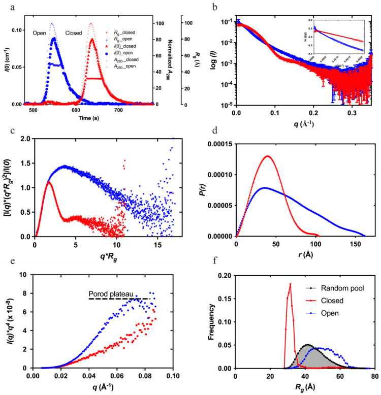
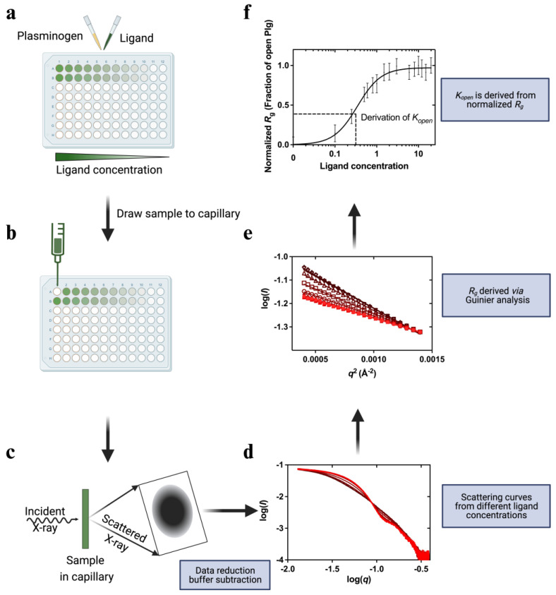
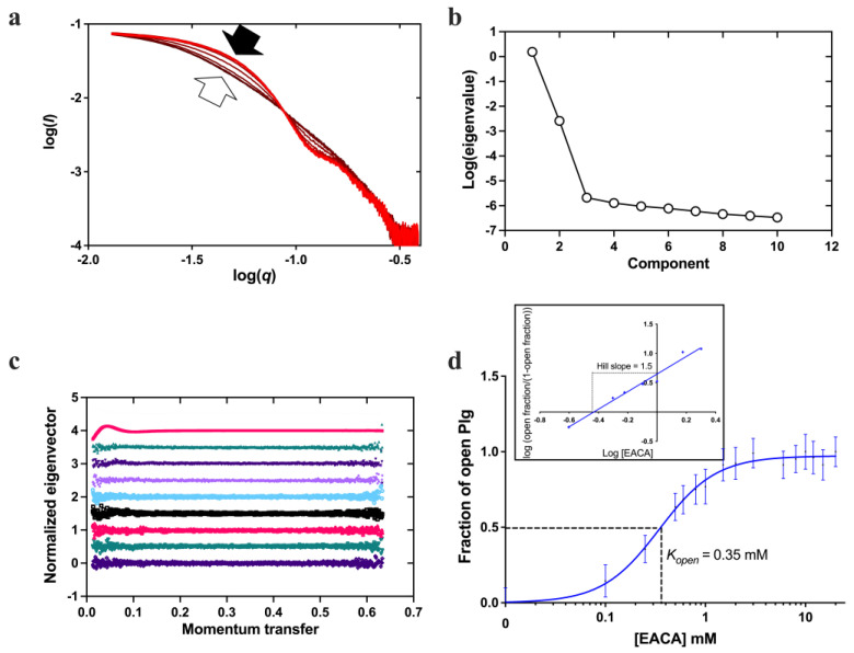
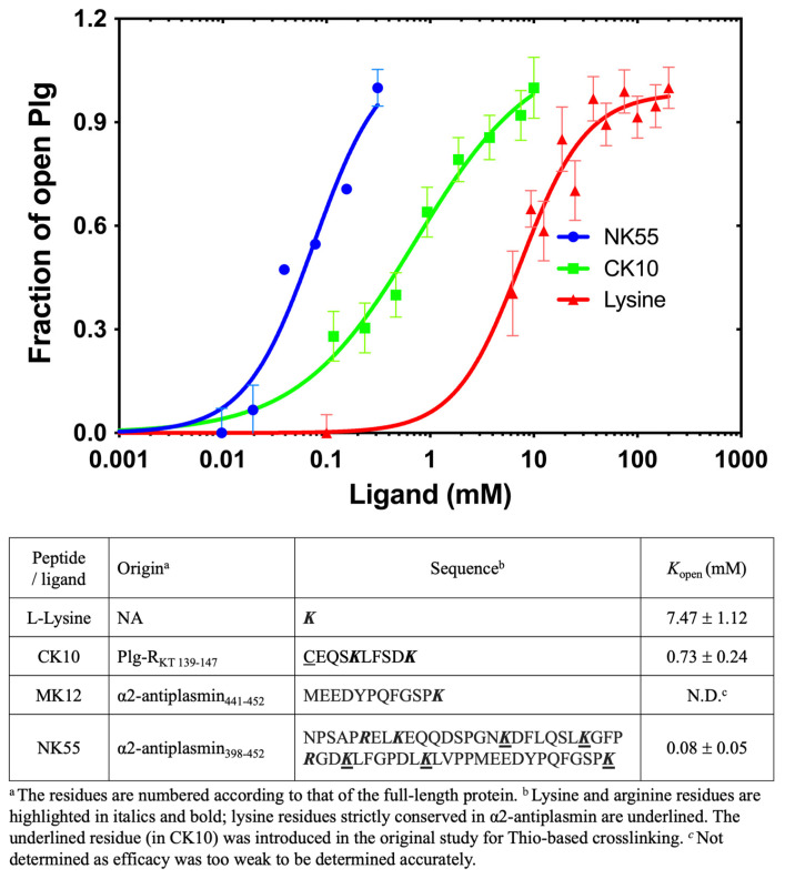
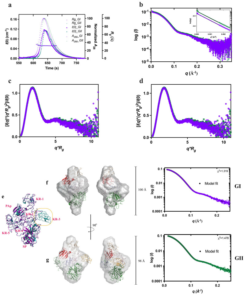
Plasminogen (Plg) is the inactive form of plasmin (Plm) that exists in two major glycoforms, referred to as glycoforms I and II (GI and GII). In the circulation, Plg assumes an activation-resistant "closed" conformation via interdomain interactions and is mediated by the lysine binding site (LBS) on the kringle (KR) domains. These inter-domain interactions can be readily disrupted when Plg binds to lysine/arginine residues on protein targets or free L-lysine and analogues. This causes Plg to convert into an "open" form, which is crucial for activation by host activators. In this study, we investigated how various ligands affect the kinetics of Plg conformational change using small-angle X-ray scattering (SAXS). We began by examining the open and closed conformations of Plg using size-exclusion chromatography (SEC) coupled with SAXS. Next, we developed a high-throughput (HTP) 96-well SAXS assay to study the conformational change of Plg. This method enables us to determine the Kopen value, which is used to directly compare the effect of different ligands on Plg conformation. Based on our analysis using Plg GII, we have found that the Kopen of ε-aminocaproic acid (EACA) is approximately three times greater than that of tranexamic acid (TXA), which is widely recognized as a highly effective ligand. We demonstrated further that Plg undergoes a conformational change when it binds to the C-terminal peptides of the inhibitor α2-antiplasmin (α2AP) and receptor Plg-RKT. Our findings suggest that in addition to the C-terminal lysine, internal lysine(s) are also necessary for the formation of open Plg. Finally, we compared the conformational changes of Plg GI and GII directly and found that the closed form of GI, which has an N-linked glycosylation, is less stable. To summarize, we have successfully determined the response of Plg to various ligand/receptor peptides by directly measuring the kinetics of its conformational changes.

Keywords: SAXS; conformational change; fibrinolysis; kringle domain; lysine analogue; lysine binding site; plasminogen; structure-function.


Conflict of interest statement

The authors declare that they have no conflict of interest with the contents of this article.

Figures

Figure 1
Figure 1
Small-angle X-ray scattering (SAXS) studies of plasminogen glycoform II (Plg GII)—showing the superposition of the closed (red) and open (blue) Plg GII: (a) coupled size-exclusion chromatography with SAXS (SEC-SAXS) profiles showing the I(0) (left Y-axis), normalized absorbance (A280), and Rg (right Y-axes); (b) SAXS profiles recorded; (c) dimensionless Kratky plot showing the globular nature of the closed and the disordered nature of the open conformation; (d) P(r) analysis shows the distinctive difference in the dimensions of the two forms; (e) Porod–Debye plots showing the Porod plateau of the closed form; and (f) ensemble optimization method (EOM) of closed and open Plg GII for analyzing the difference in Rg distribution. Distribution curves correspond to a random pool of 10,000 generated structures (grey) and the EOM-optimized ensemble of closed (blue) and open (red) Plg. The closed conformation is best represented by the compact distribution curve, and therefore, it is a homogenous ensemble. The distribution curve for the open form has shifted to the right; with a broader curve, it represents a more heterogeneous ensemble.
Figure 2
Figure 2
Overview of the high-throughput (HTP) kinetic studies of Plg conformational change. In this HTP assay, ligand-induced Plg conformational change is set up in a 96-well plate format. (a) Ligands at the study concentration are mixed with Plg and incubated for 30 min. (b,c) The samples flowed past the X-ray beam in a quartz capillary, and scattering images were collected. (d) The resulting 2D scattering profile is averaged and buffer subtracted. (e). Rg values are derived via Guinier analysis (f) and then normalized before plotting against ligand concentration. The plot is fitted to a multi-site binding model, and Kopen, the ligand concentration required to induce 50% open Plg, is derived. Kopen is a kinetic parameter indicative of ligand efficacy or stability of closed conformation.
Figure 3
Figure 3
Conformational change of Plg GII in the presence of EACA. (a) X-ray scattering curves from the EACA titration. The curves are represented in a red color gradient, from closed (light red, solid arrow) to open form (dark red, blank arrow). (b) A plot of eigenvalues from singular value decomposition (SVD) analysis of scattering curves in the EACA titration series. The number of significant eigenvalues in the plot indicates the species contributing to the scattering data. (c) A plot of successive normalized eigenvectors from SVD analysis of scattering curves (colored) in (a). The first eigenvector is displayed at the top and the last at the bottom. (d) Fractions of Plg in the open form calculated from the SAXS scattering curves are plotted against EACA concentration. A multi-site cooperative kinetic curve is fitted and shown as a solid blue line. Inset: Hill plot showing positive cooperativity mechanism (hill slope = 1.5) of EACA binding.
Figure 4
Figure 4
Conformational change of Plg GII in the presence of L-lysine and synthetic peptides derived from Plg-binding proteins. The graph shows the X-ray scattering titration curves of L-lysine (red) and synthetic peptides with a C-terminal lysine, namely CK10 (green) and NK55 (blue). MK12 was also tested, but at 10 mM, the highest concentration used, we did not observe any change in the SAXS profile of Plg GII. The Kopen derived from this study and information on the peptides regarding the corresponding residue number in the proteins and the residue sequence are shown below. Lysine and arginine residues are highlighted in bold font.
Figure 5
Figure 5
Comparison of Plg GI and GII conformations. Superposed of GI (purple) and GII (green): (a) SEC-SAXS profiles showing the I(0) (left Y-axis), normalized absorbance (A280), and Rg (right Y-axes); (b) SAXS profiles recorded; (c) dimensionless Kratky plot showing the globular nature and (d) P(r) analysis showing the similar dimensions of the two forms. (e) Superposition of Plg GI and GII crystal structures (PDB ID 4DUU and 4DUR, respectively, in the same color scheme as above). (f) Ab initio models of closed Plg GI and (g) GII; also shown are the correlation of scattering curves predicted from X-ray crystal structures using CRYSOL with the experimental data and the χ2 values.

Similar articles

Cited by

References

    1. Castellino F.J., Ploplis V.A. Structure and function of the plasminogen/plasmin system. Thromb. Haemost. 2005;93:647–654. - PubMed
    1. Rijken D.C., Lijnen H.R. New insights into the molecular mechanisms of the fibrinolytic system. J. Thromb. Haemost. January. 2009;7:4–13. doi: 10.1111/j.1538-7836.2008.03220.x. - DOI - PubMed
    1. Hayes M.L., Castellino F.J. Carbohydrate of the human plasminogen variants. II. Structure of the asparagine-linked oligosaccharide unit. J. Biol. Chem. 1979;254:8772–8776. doi: 10.1016/S0021-9258(19)86764-9. - DOI - PubMed
    1. Hayes M.L., Castellino F.J. Carbohydrate of the human plasminogen variants. III. Structure of the O-glycosidically linked oligosaccharide unit. J. Biol. Chem. 1979;254:8777–8780. doi: 10.1016/S0021-9258(19)86765-0. - DOI - PubMed
    1. Pirie-Shepherd S.R., Stevens R.D., Andon N.L., Enghild J.J., Pizzo S.V. Evidence for a novel O-linked sialylated trisaccharide on Ser-248 of human plasminogen 2. J. Biol. Chem. 1997;272:7408–7411. doi: 10.1074/jbc.272.11.7408. - DOI - PubMed

LinkOut - more resources