Review
. 2023 Sep 6;12(18):5805.
doi: 10.3390/jcm12185805.

The Heart and Seizures: Friends or Enemies?

Affiliations
Review

The Heart and Seizures: Friends or Enemies?

Elena Pasini et al. J Clin Med. .

Abstract

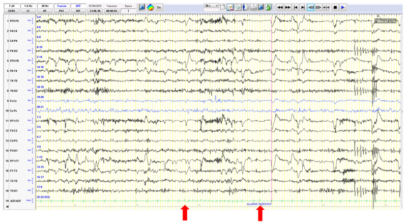
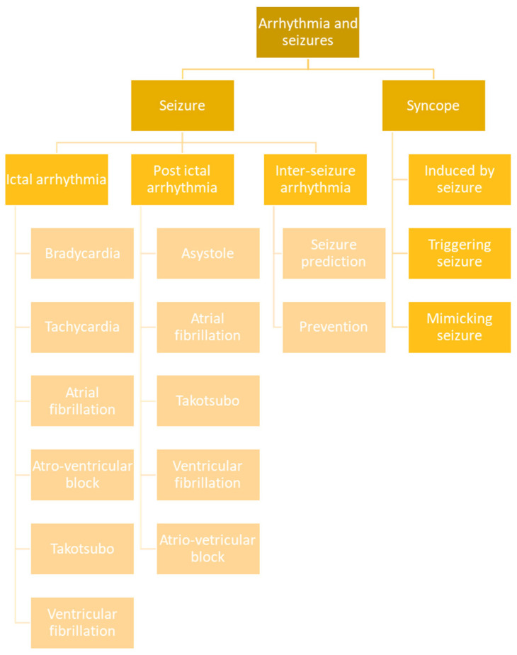
The heart and seizures are closely linked by an indissoluble relationship that finds its basis in the cerebral limbic circuit whose mechanisms remain largely obscure. The differential diagnosis between seizures and syncopes has always been a cornerstone of the collaboration between cardiologists and neurologists and is renewed as a field of great interest for multidisciplinary collaboration in the era of the diffusion of prolonged telemonitoring units. The occurrence of ictal or post-ictal arrhythmias is currently a cause of great scientific debate with respect to the role and risks that these complications can generate (including sudden unexpected death in epilepsy). Furthermore, the study of epileptic seizures and the arrhythmological complications they cause (during and after seizures) also allows us to unravel the mechanisms that link them. Finally, intercritical arrhythmias may represent great potential in terms of the prevention of cardiological risk in epileptic patients as well as in the possible prediction of the seizures themselves. In this paper, we review the pertaining literature on this subject and propose a scheme of classification of the cases of arrhythmia temporally connected to seizures.

Keywords: SUDEP; arrhythmia; epilepsy; ictal asystole; loss of consciousness; syncope.


Conflict of interest statement

The authors declare no conflict of interest.

Figures

Figure 1
Figure 1
(a): The pre-ictal HR was 80 bpm. Seizure onset is evident in the frontotemporal region with a rhythmic theta activity (see the blue arrow). HR begins to increase (see the green arrow). (b): During the same seizure (strictly confined to left frontotemporal regions and in the absence of motor involvement), HR progressively increases to 154 bpm (nearly 200% more).
Figure 2
Figure 2
Seizure onset from the left temporal region is followed (after about 8 s) by ictal asystole. During the 24 s of asystole, the EEG traces show the signs of hypoperfusion with progressive slowing of activity evolving into EEG suppression (with seizure termination after 19 s since asystole onset).
Figure 3
Figure 3
(a) The EEG traces show a right frontotemporal recruiting activity rapidly associated with ictal arrhythmia (with a 28 s asystole). (b) The right ictal activity slows down as attended in the course of hypoxic attenuation of the tracing but unexpectedly restarts over the contralateral frontotemporal areas during asystole.
Figure 4
Figure 4
ECG in Takotsubo syndrome. A 62-year-old woman affected by focal somato-sensory epilepsy with tonic–clonic evolution secondary to previous hemorrhagic stroke in the right frontal lobe. The ECG control (12 h after a cluster of seizures) showed deep anterolateral T-wave inversion and a prolonged QTc interval (493 ms). Increased troponin (689 ng/L), an echocardiogram, and coronary angiographic images confirmed the diagnosis.
Figure 5
Figure 5
EEG–EKG video recording in Brugada syndrome. The patient’s symptomatology consisted of episodes of loss of consciousness preceded by tinnitus, internal heat, chest pain, and sweating. Witnesses objectified staring and blushing followed by diffuse stiffening with urinary incontinence. See EKG changes (red arrow) with the beginning of ventricular fibrillation. The EEG only shows a secondary progressive slowdown of the background activity to theta–delta activity. The blue arrow highlights pacemaker activation.
Figure 6
Figure 6
EEG–EKG video recording in the differential diagnosis between a focal seizure and arrhythmias. A middle-aged woman complained of daily episodes of “ascending epigastric sensation”. During these symptoms, brief supraventricular tachycardia episodes (red arrows) were recorded.
Figure 7
Figure 7
Schematic representation of possible correlations between seizures and arrhythmias.

References

    1. Fong M.W.K., Norris S., Percy J., Hirsch L.J., Herlopian A. Hemisphere-Dependent Ictal Tachycardia Versus Ictal Bradycardia in a Critically Ill Patient. J. Clin. Neurophysiol. 2022;39:e15–e18. doi: 10.1097/WNP.0000000000000873. - DOI - PubMed
    1. Eggleston K.S., Olin B.D., Fisher R.S. Ictal tachycardia: The head–heart connection. Seizure. 2014;23:496–505. doi: 10.1016/j.seizure.2014.02.012. - DOI - PubMed
    1. Tinuper P., Bisulli F., Cerullo A., Carcangiu R., Marini C., Pierangeli G., Cortelli P. Ictal bradycardia in partial epileptic seizures: Autonomic investigation in three cases and literature review. Brain. 2001;124:2361–2371. doi: 10.1093/brain/124.12.2361. - DOI - PubMed
    1. Leutmezer F., Schernthaner C., Lurger S., Pötzelberger K., Baumgartner C. Electrocardiographic Changes at the Onset of Epileptic Seizures. Epilepsia. 2003;44:348–354. doi: 10.1046/j.1528-1157.2003.34702.x. - DOI - PubMed
    1. Rocamora R., Kurthen M., Lickfett L., Von Oertzen J., Elger C.E. Cardiac Asystole in Epilepsy: Clinical and Neurophysiologic Features. Epilepsia. 2003;44:179–185. doi: 10.1046/j.1528-1157.2003.15101.x. - DOI - PubMed

LinkOut - more resources