Review
. 2023 Sep 13;12(18):5949.
doi: 10.3390/jcm12185949.

Myocardial Bridging: Review on the Role of Coronary Computed Tomography Angiography

Affiliations
Review

Myocardial Bridging: Review on the Role of Coronary Computed Tomography Angiography

Chiara Rovera et al. J Clin Med. .

Abstract

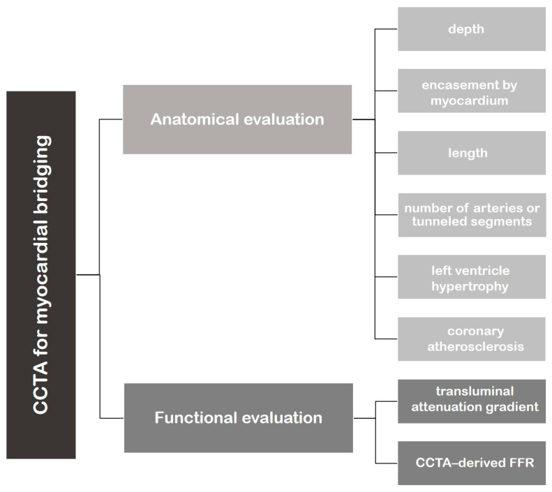
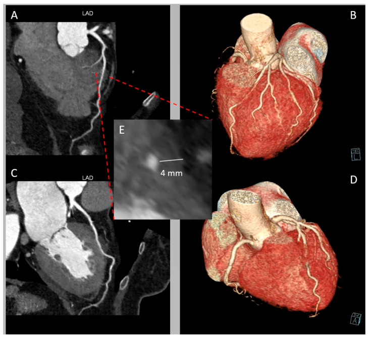
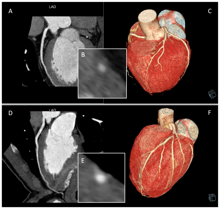
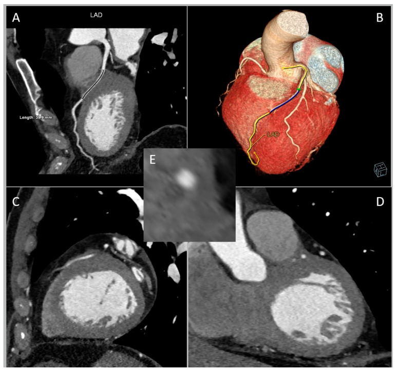
Myocardial bridging (MB) is a congenital coronary anomaly in which a segment of a coronary artery, most frequently the left anterior descending artery, deviates from its epicardial route by passing through the myocardium. The advent of cardiac computed tomography angiography (CCTA), equipped with its multiplane and three-dimensional functionalities, has notably enhanced the ability to identify MBs. Furthermore, novel post-processing methods have recently emerged to extract functional insights from anatomical evaluations. MB is generally considered a benign entity with very good survival rates; however, there is an increasing volume of evidence that certain MB characteristics may be associated with cardiovascular morbidity. This review is intended to depict the diagnostic and prognostic role of CCTA in the MB context.

Keywords: computed tomography angiography; congenital heart defects; coronary vessel anomalies; diagnostic imaging; myocardial bridging.


Conflict of interest statement

The authors declare no conflict of interest.

Figures

Figure 1
Figure 1
Flowchart representing the application of cardiac computed tomography angiography (CCTA) in the evaluation of myocardial bridging.
Figure 2
Figure 2
Deep myocardial bridging (MB) of left anterior descending artery (LAD) in a 65-year-old woman presenting with chest pain and negative V1-V5 T waves. (A) Curved multi-planar reformation of LAD providing a depiction of MB in the mid segment. (B) Whole-heart volume-rendered image showing the “tunneled” LAD. (C) Curved multi-planar reformation of LAD of the same patient in systolic phase. (D) Whole-heart volume-rendered image in systolic phase. (E) Cross-sectional image displaying LAD-MB depth.
Figure 3
Figure 3
Complete and partial myocardial bridging (MB) of left anterior descending artery (LAD). Panels (AC) are from a 54-year-old man classified as intermediate cardiovascular risk and symptomatic for chest discomfort during exertion. Panels (D)–(F) are from an 18-year-old athlete with altered cardiac repolarization during exercise. (A) Curved multi-planar reformation of LAD providing a depiction of a superficial complete MB. (B) Cross-sectional image displaying LAD-MB depth and the entire encasement by myocardium. (C) Whole-heart volume-rendered image of the same patient. (D) Curved multi-planar reformation of LAD providing a depiction of a partial MB. (E) Cross-sectional image displaying LAD-MB partial encasement. (F) Whole-heart volume-rendered image of the same patient.
Figure 4
Figure 4
Long myocardial bridging (MB) of left anterior descending artery (LAD) in a 69-year-old man referred for chest pain in arterial hypertension, dyslipidemia, and ex-smoking status. (A) Curved multi-planar reformation of LAD. (B) Whole-heart volume-rendered image. (C) Sagittal plane. (D) Coronal plane. (E) Cross-sectional image.
Figure 5
Figure 5
Schematic representation of anatomical classifications of myocardial bridging (MB).
Figure 6
Figure 6
Atherosclerotic plaque occurring proximal to the myocardial bridging (MB) of left anterior descending artery (LAD) in curved multi-planar reformation. This CCTA image belongs to an 82-year-old woman suffering from arterial hypertension and complaining of chest pain during exertion.
Figure 7
Figure 7
Deep myocardial bridging (MB) of left anterior descending artery (LAD), close to the right ventricle (RV) wall. (A) Curved multi-planar reformation of LAD. (B) Whole-heart volume-rendered image. (C) Cross-sectional image. This CCTA condition pertains to a 48-year-old woman complaining of atypical chest pain with type 2 diabetes mellitus, overweight, and smoking habit.

References

    1. Alegria J.R., Herrmann J., Holmes D.R., Jr., Lerman A., Rihal C.S. Myocardial bridging. Eur. Heart J. 2005;26:1159–1168. doi: 10.1093/eurheartj/ehi203. - DOI - PubMed
    1. Reyman H. Dissertatio de vasis cordis propriis. Diss. Ina. 1737;2:359–378.
    1. Geiringer E. The mural coronary. Am. Heart J. 1951;41:359–368. doi: 10.1016/0002-8703(51)90036-1. - DOI - PubMed
    1. Mohlenkamp S., Hort W., Ge J., Erbel R. Update on myocardial bridging. Circulation. 2002;106:2616–2622. doi: 10.1161/01.CIR.0000038420.14867.7A. - DOI - PubMed
    1. Çullu N., Yeniçeri I., Özdemir M.Y., Altun I., Doğan E. Evaluation of the morphological and clinical features of left anterior descending myocardial bridging with multi-detector computed tomography. Kardiochir Torakochirurgia Pol. 2021;18:87–91. doi: 10.5114/kitp.2021.107469. - DOI - PMC - PubMed

LinkOut - more resources