Combined Metabolic and Functional Tumor Volumes on [18F]FDG-PET/MRI in Neuroblastoma Using Voxel-Wise Analysis
- PMID: 37762918
- PMCID: PMC10531552
- DOI: 10.3390/jcm12185976
Combined Metabolic and Functional Tumor Volumes on [18F]FDG-PET/MRI in Neuroblastoma Using Voxel-Wise Analysis
Abstract
Purpose: The purpose of our study was to evaluate the association between the [18F]FDG standard uptake value (SUV) and the apparent diffusion coefficient (ADC) in neuroblastoma (NB) by voxel-wise analysis.
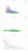
Methods: From our prospective observational PET/MRI study, a subcohort of patients diagnosed with NB with both baseline imaging and post-chemotherapy imaging was further investigated. After registration and tumor segmentation, metabolic and functional tumor volumes were calculated from the ADC and SUV values using dedicated software allowing for voxel-wise analysis. Under the mean of thresholds, each voxel was assigned to one of three virtual tissue groups: highly vital (v) (low ADC and high SUV), possibly low vital (lv) (high ADC and low SUV), and equivocal (e) with high ADC and high SUV or low ADC and low SUV. Moreover, three clusters were generated from the total tumor volumes using the method of multiple Gaussian distributions. The Pearson's correlation coefficient between the ADC and the SUV was calculated for each group.
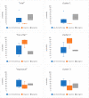
Results: Out of 43 PET/MRIs in 21 patients with NB, 16 MRIs in 8 patients met the inclusion criteria (PET/MRIs before and after chemotherapy). The proportion of tumor volumes were 26%, 36%, and 38% (v, lv, e) at baseline, 0.03%, 66%, and 34% after treatment in patients with response, and 42%, 25%, and 33% with progressive disease, respectively. In all clusters, the ADC and the SUV correlated negatively. In the cluster that corresponded to highly vital tissue, the ADC and the SUV showed a moderate negative correlation before treatment (R = -0.18; p < 0.0001) and the strongest negative correlation after treatment (R = -0.45; p < 0.0001). Interestingly, only patients with progression (n = 2) under therapy had a relevant part in this cluster post-treatment.
Conclusion: Our results indicate that voxel-wise analysis of the ADC and the SUV is feasible and can quantify the different quality of tissue in neuroblastic tumors. Monitoring ADCs as well as SUV levels can quantify tumor dynamics during therapy.
Keywords: PET/MRI; high-risk neuroblastoma; voxel-wise analysis.
Conflict of interest statement
The authors declare that they have no conflict of interest.
Figures


References
-
- Cohn S.L., Pearson A.D., London W.B., Monclair T., Ambros P.F., Brodeur G.M., Faldum A., Hero B., Iehara T., Machin D., et al. The International Neuroblastoma Risk Group (INRG) classification system: An INRG Task Force report. J. Clin. Oncol. 2009;27:289–297. doi: 10.1200/JCO.2008.16.6785. - DOI - PMC - PubMed
-
- Regier M., Derlin T., Schwarz D., Laqmani A., Henes F.O., Groth M., Buhk J.H., Kooijman H., Adam G. Diffusion weighted MRI and 18F-FDG PET/CT in non-small cell lung cancer (NSCLC): Does the apparent diffusion coefficient (ADC) correlate with tracer uptake (SUV)? Eur. J. Radiol. 2012;81:2913–2918. doi: 10.1016/j.ejrad.2011.11.050. - DOI - PubMed
-
- Monclair T., Mosseri V., Cecchetto G., De Bernardi B., Michon J., Holmes K. Influence of image-defined risk factors on the outcome of patients with localised neuroblastoma. A report from the LNESG1 study of the European International Society of Paediatric Oncology Neuroblastoma Group. Pediatr. Blood Cancer. 2015;62:1536–1542. doi: 10.1002/pbc.25460. - DOI - PubMed
LinkOut - more resources
Full Text Sources
Miscellaneous

