Review
. 2023 Sep 18;12(18):6034.
doi: 10.3390/jcm12186034.

Pedunculated Focal Nodular Hyperplasia: When in Doubt, Should We Cut It Out?

Affiliations
Review

Pedunculated Focal Nodular Hyperplasia: When in Doubt, Should We Cut It Out?

Christos Tsalikidis et al. J Clin Med. .

Abstract

Focal nodular hyperplasia (FNH) is the second most common benign hepatic tumor and can rarely present as an exophytic solitary mass attached to the liver by a stalk. Most FNH cases are usually detected as incidental findings during surgery, imaging or physical examination and have a high female predominance. However, the pedunculated forms of FNH are particularly rare and commonly associated with severe complications and diagnostic challenges. Hence, our study aims to provide a comprehensive summary of the available data on the pedunculated FNH cases among adults and children. Furthermore, we will highlight the role of different therapeutic options in treating this clinical entity. The use of imaging techniques is considered a significant addition to the diagnostic toolbox. Regarding the optimal treatment strategy, the main indications for surgery were the presence of symptoms, diagnostic uncertainty and increased risk of complications, based on the current literature. Herein, we also propose a management algorithm for patients with suspected FNH lesions. Therefore, a high index of suspicion and awareness of this pathology and its life-threatening complications, as an uncommon etiology of acute abdomen, is of utmost importance in order to achieve better clinical outcomes.

Keywords: exophytic FNH; focal nodular hyperplasia; hepatic tumor; management; pedunculated FNH.


Conflict of interest statement

The authors declare no conflict of interest.

Figures

Figure 1
Figure 1
The segmental distribution of pedunculated or exophytic FNH cases for the adult and pediatric population, based on the literature review [13,15,16,17,18,19,20,21,22,23,24,25,26,27,28,29,30,31,32,33,34,35]. IVC, inferior vena cava; GB, gallbladder; FNH, focal nodular hyperplasia.
Figure 2
Figure 2
A 31-year-old female with pedunculated FNH arising from segment III of the liver. Gross appearance of the resected and divided solitary mass with the central stellate fibrous scar (black arrow), along with an intraoperative image showing the resected exophytic mass (4.5 × 3.8 × 2.8 cm), which was attached to the liver through a rotated vascular pedicle. Preoperatively, the patient presented to the emergency department with a sudden onset of severe abdominal pain, mainly located in the epigastrium and right hypochondriac region, without any nausea, vomiting or fever. Considering the clinical manifestations and imaging findings, emergency laparoscopic resection of the tumor was successfully performed in our department. The patient data were fully anonymized without using any identifiable personal details and written informed consent was also obtained. FNH, focal nodular hyperplasia.
Figure 3
Figure 3
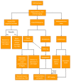
Proposed management algorithm for patients with suspected FNH. FNH, focal nodular hyperplasia; TAE, transarterial embolization; MDT, multidisciplinary team.

References

    1. Craig J.R., Peters R.L., Edmondson H.A. Atlas of Tumor Pathology. Armed Forces Institute of Pathology; Washington, DC, USA: 1989. Tumors of the Liver and Intrahepatic Bile Ducts.
    1. Wanless I.R., Mawdsley C., Adams R. On the Pathogenesis of Focal Nodular Hyperplasia of the Liver. Hepatology. 1985;5:1194–1200. doi: 10.1002/hep.1840050622. - DOI - PubMed
    1. Nahm C.B., Ng K., Lockie P. Focal Nodular Hyperplasia—A Review of Myths and Truths. J. Gastrointest. Surg. 2011;15:2275–2283. doi: 10.1007/s11605-011-1680-x. - DOI - PubMed
    1. Dioguardi B.M., Ronot M., Salvaggio G. Imaging of Hepatic Focal Nodular Hyperplasia: Pictorial Review and Diagnostic Strategy. Semin Ultrasound CT MR. 2016;37:511–524. doi: 10.1053/j.sult.2016.08.001. - DOI - PubMed
    1. Kondo F. Benign Nodular Hepatocellular Lesions Caused by Abnormal Hepatic Circulation: Etiological Analysis and Introduction of a New Concept. J. Gastroenterol. Hepatol. 2001;16:1319–1328. doi: 10.1046/j.1440-1746.2001.02576.x. - DOI - PubMed

LinkOut - more resources