. 2023 Aug 26;13(9):1812.
doi: 10.3390/life13091812.

Adiponectin Modulates Smooth Muscle Cell Morpho-Functional Properties in Murine Gastric Fundus via Sphingosine Kinase 2 Activation

Affiliations

Adiponectin Modulates Smooth Muscle Cell Morpho-Functional Properties in Murine Gastric Fundus via Sphingosine Kinase 2 Activation

Rachele Garella et al. Life (Basel). .

Abstract

Adipokines are peptide hormones produced by the adipose tissue involved in several biological functions. Among adipokines, adiponectin (ADPN) has antidiabetic and anti-inflammatory properties. It can also modulate food intake at central and peripheral levels, acting on hypothalamus and facilitating gastric relaxation. ADPN exerts its action interacting with two distinct membrane receptors and triggering some well-defined signaling cascades. The ceramidase activity of ADPN receptor has been reported in many tissues: it converts ceramide into sphingosine. In turn, sphingosine kinase (SK) phosphorylates it into sphingosine-1 phosphate (S1P), a crucial mediator of many cellular processes including contractility. Using a multidisciplinary approach that combined biochemical, electrophysiological and morphological investigations, we explored for the first time the possible role of S1P metabolism in mediating ADPN effects on the murine gastric fundus muscle layer. By using a specific pharmacological inhibitor of SK2, we showed that ADPN affects smooth muscle cell membrane properties and contractile machinery via SK2 activation in gastric fundus, adding a piece of knowledge to the action mechanisms of this hormone. These findings help to identify ADPN and its receptors as new therapeutic targets or as possible prognostic markers for diseases with altered energy balance and for pathologies with fat mass content alterations.

Keywords: adiponectin; gastric fundus; membrane properties; morphology; signaling pathway; smooth muscle; sphingosine kinase.


Conflict of interest statement

The authors declare no conflict of interest.

Figures

Figure 1
Figure 1
Adiponectin receptor 1 (AdipoR1) and 2 (AdipoR2) expression and localization. (a) Western Blot analysis performed in tissue lysates. (b,c) Representative differential interference contrast (DIC, grey) and confocal indirect immunofluorescence images of cross-section of paraffin-embedded samples stained with antibodies against AdipoR1 ((b), green) or AdipoR2 ((c), blue, pseudocolor). Nuclei are counterstained in red with Propidium iodide. Scale bar 50 μm. In (b,c): cml, circular muscle layer; lml, longitudinal muscle layer.
Figure 2
Figure 2
ADPN increases SK2 phosphorylation in murine gastric fundus. A fragment of stomach was treated with ADPN (20 nM) for 20 min before being lysed as described in the Methods Section (Section 2). Western blot analysis was performed using (a) specific anti-phospho-SK1 (Ser225) (P-SK1) and (b) anti-phospho-SK2 (Thr578) (P-SK2) antibodies. A blot representative of two independent experiments with analogous results is shown. The bar chart represents densitometric analysis of two independent experiments. Data are the mean ± SEM and are reported as protein expression normalized to GAPDH, fold change over control (set as 1). a.u.: arbitrary units. The increase in P-SK2 content induced by ADPN was found to be statistically significant by t test (* p < 0.05, treated vs. CTRL).
Figure 3
Figure 3
Involvement of SK2 in the action of ADPN in SMCs membrane properties. (a) Effect of adiponectin, ADPN (20 nM), of the sphingosine kinase 2 inhibitor SLC5111312 (SLC, 1 μM), and of SLC + ADPN on RMP values (in mV) of SMCs, compared to CTRL conditions. (p = 0.000197, one-way ANOVA with Bonferroni’s post hoc test). (b) Effect of ADPN, SLC, and SLC + ADPN on Cm (in pF) compared to CTRL condition (p = 0.00289, one-way ANOVA with Bonferroni’s post hoc test). (c) Effect of ADPN, SLC, and SLC + ADPN on Gm (in nS), compared to CTRL condition (p = 8.9 × 10⁻⁶, one-way ANOVA with Bonferroni’s post hoc test). Note the lack of effect of ADPN on passive properties of SMCs when added in the presence of SLC5111312. Data are as mean ± SD and values are listed in Table 1, together with n values. * p < 0.05 vs. CTRL; # p < 0.05 vs. ADPN.
Figure 4
Figure 4
Adiponectin (ADPN) effects on Ca2+ currents recorded from murine gastric SMCs. (a) Representative inward slowly activating Ca2+ currents (in pA) evoked with the V-clamp step protocol (inset) in untreated sample preparation (CTRL). (b) Current records from the same preparation 20 min after the application of ADPN (20 nM). (c) Overall I–V plot (with current amplitude normalized for cell capacitance, pA/pF) showing data obtained in the presence of ADPN (n = 7) and in the absence of ADPN (CTRL, n = 7). Student’s t test, * p < 0.05 vs. CTRL.
Figure 5
Figure 5
Outcome of SK2 inhibition in ADPN effects on Ca2+ ion currents recorded from murine gastric SMCs. (a) Representative current responses (in pA) evoked by the same pulse protocol reported in Figure 4a (and depicted in the inset) showing an inward slowly activating Ca2+ currents in untreated preparation (CTRL). (b) Current records from the same preparation, 20 min after the acute application of SLC (1 μM). (c) Currents recorded in the concomitant presence of SLC + ADPN. (d) I–V plot related to the currents shown in panels (ac). (e) Bar charts related to the normalized Ca2+ peak current (in pA/pF) evoked by the +10 mV step pulse. Data are mean ± SD. CTRL (n = 5), ADPN (n = 5), SLC (n = 4) and SLC + ADPN (n = 4). One way ANOVA with Bonferroni post hoc test, * p < 0.05 vs. CTRL.
Figure 6
Figure 6
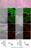
(ad) Representative images (light microscopy) of sections of paraffin-embedded muscle strips from murine gastric fundus of control (CTRL, untreated), treated with adiponectin (ADPN), or with sphingosine kinase 2 inhibitor SLC5111312 in the absence (SLC) or presence of ADPN (SLC+ ADPN) stained with hematoxylin and eosin (H&E). cml = circular muscle layer, lml = longitudinal muscle layer. (el) Representative differential interference contrast (DIC, grey) and confocal immunofluorescence images showing α-smooth muscle actin (sma) staining ((eh), green) and phospho-myosin light chain (p-MLC)2 ((il), green) Nuclei are put in evidence by PI in red. Scale bar 50 μm. (m,n) quantitative analyses of the fluorescence intensity (F.I.) in the arbitrary units (a.u.) of the indicated markers. ** p < 0.01 vs. CTRL; °° p < 0.01 vs. ADPN; ° p < 0.05 vs. ADPN (one-way ANOVA followed by Bonferroni’s post hoc test for multiple comparisons).
Figure 7
Figure 7
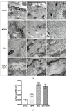
(a) Representative ultrastructural transmission electron microscopy (TEM) images of SMCs of the murine gastric fundus longitudinal muscle layer of the different indicated experimental groups. At low magnification (left panels), SMCs (nuclei labeled by asterisks) show no appreciable differences. At intermediate magnification (central panels), their cytoplasms show an extended framework of longitudinally oriented microfilaments with interposed dense bodies (DB) and converging to dense plaques (DP) at the inner aspect of the plasma membrane. In every group, the Ca2+-controlling system, made up of caveolae (C) and adjacent tubules of smooth endoplasmic reticulum (SER), and the mitochondria (M) show normal features. At high magnification (right panels), CTRL cell exhibits bundles of irregularly arranged actin contractile microfilaments; contractile filaments appear as a slightly more diffuse meshwork in ADPN-treated cell compared with the CTRL. The microfilaments appear to form a looser network in the SLC- and SLC + ADPN-treated cells. (b) The bar charts show the morphometrical analysis of relative area of the microfilament framework, evaluated on 20 regions of interest (ROI, 40,820 nm2 each) in high-magnification ultrastructural pictures of longitudinally sectioned SMC from the different experimental groups. * p < 0.05, *** p < 0.001 vs. CTRL (one-way ANOVA and Newman–Keuls multiple comparison post test).

References

    1. Rozwadowski J., Borodzicz-Jażdżyk S., Czarzasta K., Cudnoch-Jędrzejewska A. A Review of the Roles of Apelin and ELABELA Peptide Ligands in Cardiovascular Disease, Including Heart Failure and Hypertension. Med. Sci. Monit. 2022;28:e938112. doi: 10.12659/MSM.938112. - DOI - PMC - PubMed
    1. Su X., Zhang G., Cheng Y., Wang B. Leptin in skin disease modulation. Clin. Chim. Acta. 2021;516:8–14. doi: 10.1016/j.cca.2021.01.013. - DOI - PubMed
    1. Schüler-Toprak S., Ortmann O., Buechler C., Treeck O. The Complex Roles of Adipokines in Polycystic Ovary Syndrome and Endometriosis. Biomedicines. 2022;10:2503. doi: 10.3390/biomedicines10102503. - DOI - PMC - PubMed
    1. Schmitt M., Gallistl J., Schüler-Toprak S., Fritsch J., Buechler C., Ortmann O., Treeck O. Anti-Tumoral Effect of Chemerin on Ovarian Cancer Cell Lines Mediated by Activation of Interferon Alpha Response. Cancers. 2022;14:4108. doi: 10.3390/cancers14174108. - DOI - PMC - PubMed
    1. Shklyaev S.S., Melnichenko G.A., Volevodz N.N., Falaleeva N.A., Ivanov S.A., Kaprin A.D., Mokrysheva N.G. Adiponectin: A pleiotropic hormone with multifaceted roles. Probl. Endocrinol. 2021;67:98–112. doi: 10.14341/probl12827. - DOI - PMC - PubMed

LinkOut - more resources