Review
. 2023 Aug 24;59(9):1530.
doi: 10.3390/medicina59091530.

Indocyanine Green (ICG) and Colorectal Surgery: A Literature Review on Qualitative and Quantitative Methods of Usage

Affiliations
Review

Indocyanine Green (ICG) and Colorectal Surgery: A Literature Review on Qualitative and Quantitative Methods of Usage

Laurentiu Simion et al. Medicina (Kaunas). .

Abstract

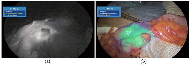
Background: Due to its many benefits, indocyanine green (ICG) has gained progressive popularity in operating rooms (ORs) globally. This literature review examines its qualitative and quantitative usage in surgical treatment. Method: Relevant terms were searched in five international databases (1. Pubmed, 2. Sciencedirect, 3. Scopus, 4. Oxfordjournals, 5. Reaxys) for a comprehensive literature review. The main benefits of using ICG in colorectal surgery are: intraoperative fluorescence angiography; fluorescence-guided lymph node involvement detection and the sentinel technique; the fluorescent emphasis of a minute liver tumour, counting just 200 tumour cells; facilitation of fistula diagnosis; and tumour tattooing. This methodology can also be used with quantitative characteristics such as maximum intensity, relative maximum intensity, and in-flow parameters such as time-to-peak, slope, and t1/2max. This article concludes that fluorescence surgery with ICG and near-infrared (NIR) light is a relatively new technology that improves anatomical and functional information, allowing more comprehensive and safer tumour removal and the preservation of important structures.

Keywords: ICG; ICG-NIR; colorectal; colorectal surgery; fluorescence; intraoperative staining; q-ICG.


Conflict of interest statement

The authors declare no conflict of interest, except the fact that two previous (extensively modified) versions of this article were published as Preprints and can be found at the following address: https://www.preprints.org/manuscript/202205.0383/v1, accessed on 20 August 2023.

Figures

Figure 1
Figure 1
a and 1.b. ICG-enhanced fluorescence-guided evaluation of colonic perfusion following division of the mesentery during laparoscopic left colectomy. Intraoperative Monochromatic NIR/ICG (a) and Intensity Map (b) images. The forceps tip marks the well-perfused area.
Figure 2
Figure 2
In this situation, ICG-enhanced fluorescence imaging helps locate metastatic tumours and determine resection margins. Figure 2 Enhanced fluorescence-guided liver resection for metastatic hepatic lesions (with ICG). The intraoperative images (ag) were captured using the following visualisation modes: (a) White light mode, (b) NIR/ICG Overlay mode, (c) NIR/ICG Intensity Map mode, (d) NIR/ICG Overlay mode (open surgery), (e) NIR/ICG Overlay mode (Macroscopic image of surgical specimen following extraction), (f) NIR/ICG Monochromatic mode of same specimen as in (e), and (g) NIR/ICG Intensity Map mode showing the same specimen as in (e).
Figure 2
Figure 2
In this situation, ICG-enhanced fluorescence imaging helps locate metastatic tumours and determine resection margins. Figure 2 Enhanced fluorescence-guided liver resection for metastatic hepatic lesions (with ICG). The intraoperative images (ag) were captured using the following visualisation modes: (a) White light mode, (b) NIR/ICG Overlay mode, (c) NIR/ICG Intensity Map mode, (d) NIR/ICG Overlay mode (open surgery), (e) NIR/ICG Overlay mode (Macroscopic image of surgical specimen following extraction), (f) NIR/ICG Monochromatic mode of same specimen as in (e), and (g) NIR/ICG Intensity Map mode showing the same specimen as in (e).
Figure 3
Figure 3
Left ureter fluorescence imaging during left colectomy with ICG: (a,b) intraoperative pictures from green NIR/ICG Overlay mode.

References

    1. Ottobrini L., Martelli C., Lucignani G. Optical Imaging Agents. Mol. Imaging. 2021:603–625. doi: 10.1016/B978-0-12-816386-3.00035-1. - DOI
    1. Pirovano G., Roberts S., Kossatz S., Reiner T. Optical Imaging Modalities: Principles and Applications in Preclinical Research and Clinical Settings. J. Nucl. Med. 2020;61:1419–1427. doi: 10.2967/jnumed.119.238279. - DOI - PubMed
    1. Yang L., Huang B., Hu S., An Y., Li Y., Wang Y., Gu N. Indocyanine green assembled free oxygen-nanobubbles towards enhanced near-infrared induced photodynamic therapy. Nano Res. 2022;15:4285–4293. doi: 10.1007/s12274-022-4085-0. - DOI - PMC - PubMed
    1. Lee E.H., Lee M.K., Lim S.J. Enhanced stability of indocyanine green by encapsulation in zein-phosphatidylcholine hybrid nanoparticles for use in the phototherapy of cancer. Pharmaceutics. 2021;13:305. doi: 10.3390/pharmaceutics13030305. - DOI - PMC - PubMed
    1. Qing W., Xing X., Feng D., Chen R., Liu Z. Indocyanine green loaded pH-responsive bortezomib supramolecular hydrogel for synergistic chemo-photothermal/photodynamic colorectal cancer therapy. Photodiagn. Photodyn. Ther. 2021;36:102521. doi: 10.1016/j.pdpdt.2021.102521. - DOI - PubMed

LinkOut - more resources