Review
. 2023 Sep 2;59(9):1589.
doi: 10.3390/medicina59091589.

Image-Guided Prostate Cryoablation: State-of-the-Art

Affiliations
Review

Image-Guided Prostate Cryoablation: State-of-the-Art

Vijay Ramalingam et al. Medicina (Kaunas). .

Abstract

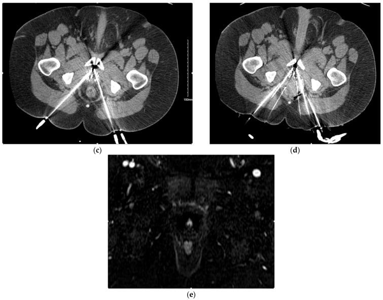
Image-guided focal therapy has increased in popularity as a treatment option for patients with primary and locally recurrent prostate cancer. This review will cover the basic indications, evaluation, treatment algorithm, and follow-up for patients undergoing image-guided ablation of the prostate. Additionally, this paper will serve as an overview of some technical approaches to cases so that physicians can familiarize themselves with working in this space. While the focus of this paper is prostate cryoablation, readers will obtain a basic literature overview of some of the additional available image-guided treatment modalities for focal prostate therapy.

Keywords: men’s health; prostate cryoablation; prostate focal therapy.


Conflict of interest statement

There are no conflict of interest to declare from any participating author.

Figures

Figure 1
Figure 1
Case example of focal CT-guided cryoablation of locally recurrent prostate cancer. (a) Fused axial images of a fluciclovine-flourine-18-labeled SPECT/CT showing a focus of avidity along the right peripheral zone apex, correlating with local prostate cancer recurrence. (b) Axial contrast-enhanced T1-weighted MRI subtraction image showing the enhancement of locally recurrent prostate cancer within the right peripheral zone apex correlating with area of avidity (a). (c) Axial CT images showing transgluteal IcePearl cryoablation needle within the target lesion within the right hemi-prostate and placement of perirectal hydrodissection needle. (d) Transgluteal cryoablation probe into the prostate target lesion after instillation of 2% contrast:0.9% saline solution within the perirectal region to safely separate critical structures for cryoablation. (e) Fused axial images of the post-ablation 68Ga-PSMA-11 PET-CT showing no residual avidity along the right peripheral zone apex.
Figure 1
Figure 1
Case example of focal CT-guided cryoablation of locally recurrent prostate cancer. (a) Fused axial images of a fluciclovine-flourine-18-labeled SPECT/CT showing a focus of avidity along the right peripheral zone apex, correlating with local prostate cancer recurrence. (b) Axial contrast-enhanced T1-weighted MRI subtraction image showing the enhancement of locally recurrent prostate cancer within the right peripheral zone apex correlating with area of avidity (a). (c) Axial CT images showing transgluteal IcePearl cryoablation needle within the target lesion within the right hemi-prostate and placement of perirectal hydrodissection needle. (d) Transgluteal cryoablation probe into the prostate target lesion after instillation of 2% contrast:0.9% saline solution within the perirectal region to safely separate critical structures for cryoablation. (e) Fused axial images of the post-ablation 68Ga-PSMA-11 PET-CT showing no residual avidity along the right peripheral zone apex.
Figure 2
Figure 2
Case example of focal CT-guided cryoablation of locally recurrent prostate cancer. (a) Fused axial images of a fluciclovine-flourine-18-labeled SPECT/CT showing a focus of avidity along the midline anterior mid-gland, correlating with local prostate cancer recurrence. (b) Axial contrast-enhanced T1-weighted MRI subtraction image showing the enhancement of locally recurrent prostate cancer within the midline anterior mid-gland correlating with area of avidity (a). (c) Axial CT images showing multiple transgluteal IcePearl cryoablation needles within the target lesion within the midline anterior mid-gland. (d) Transgluteal cryoablation probe into the prostate target lesion after instillation of 2% contrast:0.9% saline solution within the perirectal region to safely separate critical structures for cryoablation after placement of multiple hydrodissection needles; images also demonstrate ice ball formation within the target lesion. (e) Axial contrast-enhanced T1-weighted MRI subtraction image showing resolution of enhancement along the targeted region post-ablation.
Figure 2
Figure 2
Case example of focal CT-guided cryoablation of locally recurrent prostate cancer. (a) Fused axial images of a fluciclovine-flourine-18-labeled SPECT/CT showing a focus of avidity along the midline anterior mid-gland, correlating with local prostate cancer recurrence. (b) Axial contrast-enhanced T1-weighted MRI subtraction image showing the enhancement of locally recurrent prostate cancer within the midline anterior mid-gland correlating with area of avidity (a). (c) Axial CT images showing multiple transgluteal IcePearl cryoablation needles within the target lesion within the midline anterior mid-gland. (d) Transgluteal cryoablation probe into the prostate target lesion after instillation of 2% contrast:0.9% saline solution within the perirectal region to safely separate critical structures for cryoablation after placement of multiple hydrodissection needles; images also demonstrate ice ball formation within the target lesion. (e) Axial contrast-enhanced T1-weighted MRI subtraction image showing resolution of enhancement along the targeted region post-ablation.

References

    1. Culp M.B., Soerjomataram I., Efstathiou J.A., Bray F., Jemal A. Recent Global Patterns in Prostate Cancer Incidence and Mortality Rates. Eur. Urol. 2020;77:38–52. doi: 10.1016/j.eururo.2019.08.005. - DOI - PubMed
    1. Cooperberg M.R., Lubeck D.P., Meng M.V., Mehta S.S., Carroll P.R. The changing face of low-risk prostate cancer: Trends in clinical presentation and primary management. J. Clin. Oncol. 2004;22:2141–2149. doi: 10.1200/JCO.2004.10.062. - DOI - PMC - PubMed
    1. Hamdy F.C., Donovan J.L., Lane J.A., Metcalfe C., Davis M., Turner E.L., Martin R.M., Young G.J., Walsh E.I., Bryant R.J., et al. Fifteen-Year Outcomes after Monitoring, Surgery, or Radiotherapy for Prostate Cancer. N. Engl. J. Med. 2023;388:1547–1558. doi: 10.1056/NEJMoa2214122. - DOI - PubMed
    1. Lei J.H., Liu L.R., Wei Q., Yan S.B., Song T.R., Lin F.S., Yang L., Cao D.H., Yuan H.C., Xue W.B., et al. Systematic review and meta-analysis of the survival outcomes of first-line treatment options in high-risk prostate cancer. Sci. Rep. 2015;5:7713. doi: 10.1038/srep07713. - DOI - PMC - PubMed
    1. Petrelli F., Vavassori I., Coinu A., Borgonovo K., Sarti E., Barni S. Radical prostatectomy or radiotherapy in high-risk prostate cancer: A systematic review and metaanalysis. Clin. Genitourin. Cancer. 2014;12:215–224. doi: 10.1016/j.clgc.2014.01.010. - DOI - PubMed

LinkOut - more resources