Review
. 2023 Sep 7;14(9):1744.
doi: 10.3390/mi14091744.

Point-of-Care Devices for Viral Detection: COVID-19 Pandemic and Beyond

Affiliations
Review

Point-of-Care Devices for Viral Detection: COVID-19 Pandemic and Beyond

Sumit K Yadav et al. Micromachines (Basel). .

Abstract

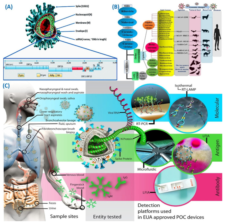
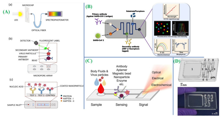
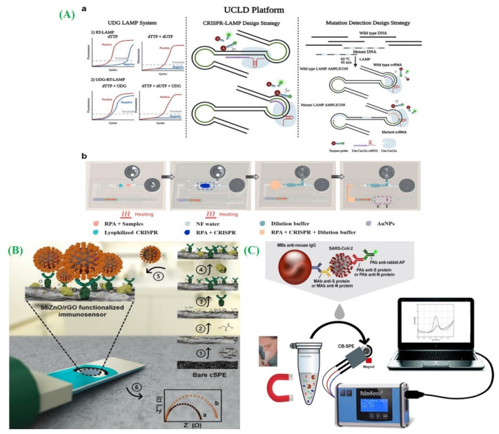
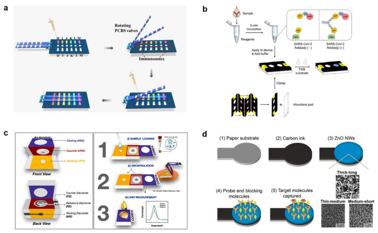
The pandemic of COVID-19 and its widespread transmission have made us realize the importance of early, quick diagnostic tests for facilitating effective cure and management. The primary obstacles encountered were accurately distinguishing COVID-19 from other illnesses including the flu, common cold, etc. While the polymerase chain reaction technique is a robust technique for the determination of SARS-CoV-2 in patients of COVID-19, there arises a high demand for affordable, quick, user-friendly, and precise point-of-care (POC) diagnostic in therapeutic settings. The necessity for available tests with rapid outcomes spurred the advancement of POC tests that are characterized by speed, automation, and high precision and accuracy. Paper-based POC devices have gained increasing interest in recent years because of rapid, low-cost detection without requiring external instruments. At present, microfluidic paper-based analysis devices have garnered public attention and accelerated the development of such POCT for efficient multistep assays. In the current review, our focus will be on the fabrication of detection modules for SARS-CoV-2. Here, we have included a discussion on various strategies for the detection of viral moieties. The compilation of these strategies would offer comprehensive insight into the detection of the causative agent preparedness for future pandemics. We also provide a descriptive outline for paper-based diagnostic platforms, involving the determination mechanisms, as well as a commercial kit for COVID-19 as well as their outlook.

Keywords: COVID-19; POC testing devices; SARS-CoV-2; diagnostics; viral sensor.


Conflict of interest statement

The authors declare no conflict of interest.

Figures

Figure 1
Figure 1
(A) Scheme depicting the structure of the SARS-CoV-2 virus (adapted with permission from Ref. [55], copyright 2020 MDPI). (B) Coronavirus taxonomy as per ICTV displaying the SARS-CoV-2 classification (adapted with permission from Ref. [56], copyright 2020 MDPI). (C) Human samples summary in which SARS-CoV-2 virus could be identified targets could be investigated, and detection platforms used in EUA-approved POC devices (adapted with permission from Ref. [48], copyright 2021 American Chemical Society).
Figure 3
Figure 3
(A) Optical-based microfluidic platforms have been illustrated for the detection of respiratory viruses or their products using various techniques: (a) detection based on absorbance measurements, (b) fluorescent detection utilizing a micropore array, and (c) colorimetric detection employing a paper-based microfluidic platform with RT-LAMP (adapted with permission from Ref. [73], copyright 2021 Springer Nature). (B) Several microfluidics techniques have shown potential for the detection of SARS-CoV-2 using antibodies against the virus (adapted with permission from Ref. [69], copyright 2020 Frontiers). (C) Schematic illustration of an overview of several microfluidic techniques used for respiratory virus detection based on nanoparticles, aptamers, enzymes, antibodies, or magnetic beads (adapted with permission from Ref. [73], copyright 2021 Springer Nature). (D) The IFAST RT-LAMP device is designed with a sample chamber that is interconnected to wash chambers and a detection chamber through gates and a photograph of an IFAST RT-LAMP microfluidic device specifically developed for the detection of SARS-CoV-2 RNA (adapted with permission from Ref. [72], copyright 2021 Elsevier).
Figure 6
Figure 6
(A) An overview of the CRISPR–LAMP detection platform, designed to offer universal stability and precise results (a) (adapted with permission from Ref. [89], copyright 2020 American Chemical Society), and a microfluidic device utilizing lateral flow technology, which operates independently without the need for external devices, and incorporates a hand-warmer pouch as its power source (b) (adapted with permission from Ref. [92], copyright 2022 Elsevier); (B) a graphical representation depicting the sequential process of immuno-biosensor preparation (adapted with permission from Ref. [101], copyright 2020 American Chemical Society); and (C) a schematic demonstration of the detection process of electrochemical biosensor for a saliva sample (adapted with permission from Ref. [103], copyright 2020 Elsevier).
Figure 10
Figure 10
(a) PFST-μPADs for quantitative SARS-CoV-2 IgA/IgM/IgG assay (adapted with permission from Ref. [136], copyright 2021 American Chemical Society). (b) 3D μPADs are utilized for the detection of SARS-CoV-2 specific antibodies by leveraging the affinity between cellulose and the cellulose binding domain (adapted with permission from Ref. [139], copyright 2021 American Chemical Society). (c) A label-free ePAD for detecting SARS-CoV-2-specifc IgG and IgM antibodies (adapted with permission from Ref. [70], copyright 2021 Elsevier). (d) A novel ePAD proposed for COVID-19 diagnosis, incorporating a working electrode enhanced by ZnO nanowires (adapted with permission from Ref. [142], copyright 2021 Elsevier).
Figure 2
Figure 2
(i) (A) The microfluidic chip design comprises 1024-unit cells, enabling high-throughput detection of SARS-CoV-2. (B) The experimental process is depicted in an illustrative manner. (C) The chip facilitates the sandwich immunoassay process. (D) The fluorescence response of antihuman IgG-PE is observed for the anti-spike antibodies present in human serum. (E) An image displays the limit of detection (LOD) marked with a dashed line, along with the concentration of antihuman IgG-PE against anti-spike IgG. (ii) Schematic illustration of ultralow-volume whole blood sampling and processing developed by Swank et al. (A) The Mitra® device and (B) the HemaXis™DB10 device are used to collect 10 μL of whole blood, while (C) the blood glucose test strip collects 0.6 μL of whole blood. After collection, (DF) the blood samples are dried, allowing the devices to be shipped via regular mail under ambient conditions. Upon arrival at the laboratory, (G) the Mitra® tips are removed and placed in a 96-well plate, (H) the HemaXis™DB10 cards are punched, and the filter discs are placed in a 96-well plate, and (I) the glucose test strip is cut to size and placed in an Eppendorf tube. Next, (JL) the blood samples are extracted in a buffer solution through overnight incubation at 4 °C, followed by (M) transfer to a spotting plate. Subsequently, the samples are (N) microarrayed and (O) analyzed using the NIA device (adapted with permission from Ref. [68], copyright 2021 Proceedings of the National Academy of Sciences).
Figure 4
Figure 4
(A) A schematic diagram illustrating the process of LAMP detection (adapted with permission from Ref. [74], copyright 2020 MDPI). (B) The microfluidic chip is designed with a sequential fluid dispensing structure (adapted with permission from Ref. [75], copyright 2021 Royal Society of Chemistry). (C) Illustration of digital LAMP quantification having the da-Slip Chip by utilizing the random-access system (adapted with permission from Ref. [76], copyright 2021 Royal Society of Chemistry). (D) A schematic demonstration depicting the process of RT-LAMP amplification and detection in centrifugal PS-T microdevices: (i) introduction of reagents and sealing with transparent contact paper; (ii) incubation in a thermo block; (iii) centrifugation using a fidget spinner to rupture the valve; and (iv) visual detection under UV radiation (adapted with permission from Ref. [77], copyright 2021 Royal Society of Chemistry). (E) Development of a LAMP-integrated microfluidic chip and particle diffusometry (adapted with permission from Ref. [78], copyright 2022 Elsevier).
Figure 5
Figure 5
(a) A photograph showcasing a compact and portable centrifugal microfluidic device; (b) a schematic diagram illustrating the different components of the microfluidic device; and (c) a schematic illustration highlighting the structure and functional regions of the centrifugal microfluidic chip (adapted with permission from Ref. [83], copyright 2020 Royal Society of Chemistry).
Figure 7
Figure 7
(A) Cellular receptor (ACE2)-based LFA for detecting SARS-CoV-2 S1 antigen (adapted with permission from Ref. [122], copyright 2021 Elsevier). (B) Development of highly specific and sensitive scFv-Fc fusion proteins (rapidly screened by phage display technology) based LFA for detection of the SARS-CoV-2 N protein (adapted with permission from Ref. [124], copyright 2021 Elsevier).
Figure 8
Figure 8
(A) The detection system is configured to quantify the results of the LFA using a photon-counting approach for the detection of IgG antibodies (adapted with permission from Ref. [129], copyright 2020 American Institute of Physics); and (B) the LFA strip designed to detect anti-SARS-CoV-2 IgA antibodies and a simple and universally compatible smartphone reader is employed to detect the optical signal emitted from the LFA (adapted with permission from Ref. [130], copyright 2021 Elsevier).
Figure 9
Figure 9
(A) The lateral flow strip membranes (LFSM) for the highly specific and sensitive method for detecting SARS-CoV-2 that enable simultaneous detection of multiple regions of SARS-CoV-2 RNA in a single test (adapted with permission from Ref. [131,132], copyright 2020 American Chemical Society). (B) The reverse transcription-enzymatic recombinase amplification (RT-ERA) operates on the principle of ultrasensitive, field-deployable, and simultaneous dual-gene detection of SARS-CoV-2 RNA (adapted with permission from Ref. [134], copyright 2020 Springer Nature).

Similar articles

Cited by

References

    1. Mishra A., Nair N., Yadav A.K., Solanki P., Majeed J., Tripathi V. SARS-CoV-2 Origin and COVID-19 Pandemic Across the Globe. Volume 41 IntechOpen; London, UK: 2021. Coronavirus Disease 2019 (COVID-19): Origin, Impact, and Drug Development.
    1. Verma D., Yadav A.K., Chaudhary N., Mukherjee M.D., Kumar P., Kumar A., Solanki P.R. Recent Advances in Understanding SARS-CoV-2 Infection and Updates on Potential Diagnostic and Therapeutics for COVID-19. Coronaviruses. 2022;3:14–31.
    1. Mofijur M., Fattah I.M.R., Alam M.A., Islam A.B.M.S., Ong H.C., Rahman S.M.A., Najafi G., Ahmed S.F., Uddin M.A., Mahlia T.M.I. Impact of COVID-19 on the social, economic, environmental and energy domains: Lessons learnt from a global pandemic. Sustain. Prod. Consum. 2021;26:343–359. doi: 10.1016/j.spc.2020.10.016. - DOI - PMC - PubMed
    1. Sohrabi C., Alsafi Z., O’neill N., Khan M., Kerwan A., Al-Jabir A., Iosifidis C., Agha R. World Health Organization declares global emergency: A review of the 2019 novel coronavirus (COVID-19) Int. J. Surg. 2020;76:71–76. doi: 10.1016/j.ijsu.2020.02.034. - DOI - PMC - PubMed
    1. Wang C., Wu J., He Y., Song Z., Shi S., Zhu Y., Jia Y., Ye W. Fully solid-state graphene transistors with striking homogeneity and sensitivity for the practicalization of single-device electronic bioassays. Nano Lett. 2019;20:166–175. doi: 10.1021/acs.nanolett.9b03528. - DOI - PubMed