

A Multi-Layer Breast Cancer Model to Study the Synergistic Effect of Photochemotherapy
- PMID: 37763969
- PMCID: PMC10535669
- DOI: 10.3390/mi14091806
A Multi-Layer Breast Cancer Model to Study the Synergistic Effect of Photochemotherapy
Abstract
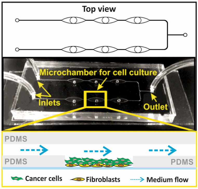
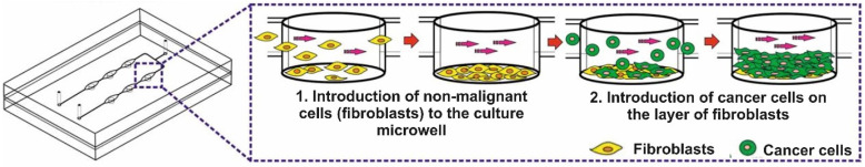
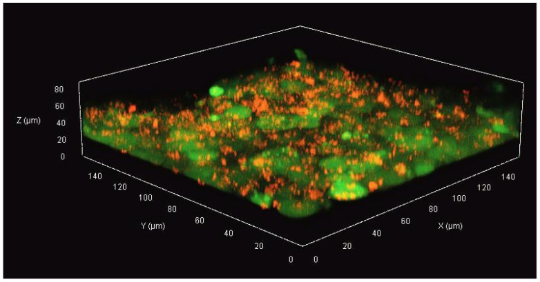
Breast cancer is one of the most common cancers among women. The development of new and effective therapeutic approaches in the treatment of breast cancer is an important challenge in modern oncology. Two-dimensional (2D) cell cultures are most often used in the study of compounds with potential anti-tumor nature. However, it is necessary to develop advanced three-dimensional (3D) cell models that can, to some extent, reflect the physiological conditions. The use of miniature cancer-on-a-chip microfluidic systems can help to mimic the complex cancer microenvironment. In this report, we developed a 3D breast cancer model in the form of a cell multilayer, composed of stromal cells (HMF) and breast cancer parenchyma (MCF-7). The developed cell model was successfully used to analyze the effectiveness of combined sequential photochemotherapy, based on doxorubicin and meso-tetraphenylporphyrin. We proved that the key factor that allows achieving the synergistic effect of combination therapy are the order of drug administration to the cells and the sequence of therapeutic procedures. To the best of our knowledge, studies on the effectiveness of combination photochemotherapy depending on the sequence of the component drugs were performed for the first time under microfluidic conditions on a 3D multilayered model of breast cancer tissue.
Keywords: breast cancer; cancer-on-a-chip; meso-tetraphenylporphyrin; multilayered cell model; sequential photochemotherapy; three-dimensional (3D) cell culture.
Conflict of interest statement
The authors declare no conflict of interest.
Figures
References
-
- Cancer. [(accessed on 23 August 2023)]. Available online: https://www.who.int/health-topics/cancer.
-
- Mota A., Colás E., García-Sanz P., Campoy I., Rojo-Sebastián A., Gatius S., García Á., Chiva L., Alonso S., Gil-Moreno A., et al. Genetic Analysis of Uterine Aspirates Improves the Diagnostic Value and Captures the Intra-Tumor Heterogeneity of Endometrial Cancers. Mod. Pathol. 2017;30:134–145. doi: 10.1038/modpathol.2016.143. - DOI - PubMed
-
- Phipps A.I., Li C.I., Kerlikowske K., Barlow W.E., Buist D.S.M. Risk Factors for Ductal, Lobular, and Mixed Ductal-Lobular Breast Cancer in a Screening Population. Cancer Epidemiol. Biomark. Prev. Publ. Am. Assoc. Cancer Res. Cosponsored Am. Soc. Prev. Oncol. 2010;19:1643–1654. doi: 10.1158/1055-9965.EPI-10-0188. - DOI - PMC - PubMed
LinkOut - more resources
Full Text Sources
