. 2023 Sep 7;28(18):6505.
doi: 10.3390/molecules28186505.

Response of Osteoblasts on Amine-Based Nanocoatings Correlates with the Amino Group Density

Affiliations

Response of Osteoblasts on Amine-Based Nanocoatings Correlates with the Amino Group Density

Susanne Seemann et al. Molecules. .

Abstract

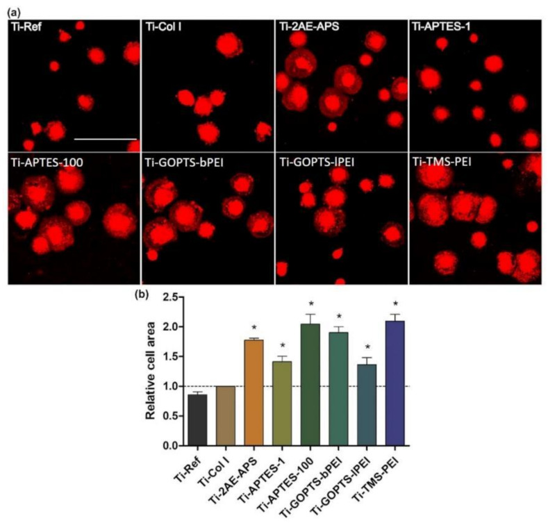
Increased life expectancy in industrialized countries is causing an increased incidence of osteoporosis and the need for bioactive bone implants. The integration of implants can be improved physically, but mainly by chemical modifications of the material surface. It was recognized that amino-group-containing coatings improved cell attachment and intracellular signaling. The aim of this study was to determine the role of the amino group density in this positive cell behavior by developing controlled amino-rich nanolayers. This work used covalent grafting of polymer-based nanocoatings with different amino group densities. Titanium coated with the positively-charged trimethoxysilylpropyl modified poly(ethyleneimine) (Ti-TMS-PEI), which mostly improved cell area after 30 min, possessed the highest amino group density with an N/C of 32%. Interestingly, changes in adhesion-related genes on Ti-TMS-PEI could be seen after 4 h. The mRNA microarray data showed a premature transition of the MG-63 cells into the beginning differentiation phase after 24 h indicating Ti-TMS-PEI as a supportive factor for osseointegration. This amino-rich nanolayer also induced higher bovine serum albumin protein adsorption and caused the cells to migrate slower on the surface after a more extended period of cell settlement as an indication of a better surface anchorage. In conclusion, the cell spreading on amine-based nanocoatings correlated well with the amino group density (N/C).

Keywords: actin cytoskeleton; amino coating; gene expression; material characterization; microarray; polymer; spreading; surface charge; surface modification; wettability.


Conflict of interest statement

The authors declare no conflict of interest.

Figures

Figure 1
Figure 1
Spreading capacity of MG-63s on amine-based coatings after 30 min. (a) Cell area of membrane-stained cells (red). Note that cell areas increased on all amino-rich nanolayers compared to pure Ti-Ref and the extracellular matrix protein Col I as controls. (confocal microscopy, LSM 780, Carl Zeiss; red: PKH-26; scale bar 100 μm; abbreviations see Table 1). (b) Relative cell areas—the spreading values were compared to Col I (mean ± s.e.m.; n = 3 independent experiments with 50 cells each; unpaired t-test; * p < 0.05).
Figure 2
Figure 2
Actin cytoskeleton of MG-63 cells on amine-based Ti-TMS-PEI vs. uncoated Ti. (a) Cell spreading and development of actin filaments in the time frame 4 h and 24 h on planar Ti samples. MG-63s showed an increased cell area on Ti-TMS-PEI after 4 h compared to Ti-Ref, and the actin filaments are more pronounced. After 24 h, these differences in cell size and filament organization are equalized (representative figures of n = 3 independent experiments). (b) Cell orientation and actin formation of MG-63s on micro-grooved Ti-G20 structures after 24 h. Cells on grooved Ti-Ref samples (20 µm in width, 2 µm in height) appear aligned due to contact guidance. In contrast, cells on grooved Ti-TMS-PEI experience a slight abrogation of the contact guidance, i.e., the nanolayer has an impact that is dominant over the micro-structure (3D z-stack, dashed lines indicating groove edges, representative figures of n = 2 independent experiments). (For all images: confocal microscopy, LSM 780, Carl Zeiss; actin: phalloidin-TRITC, red, nucleus: DAPI, blue; scale bars 10 μm).
Figure 3
Figure 3
Morphology of MG-63 osteoblast-like cells on TMS-PEI-coated Ti for 30 min and 24 h versus the pure Ti-Ref. Note the considerably enhanced cell area after 30 min. No morphological differences are evident after 24 h (representative images of n = 3 independent experiments, scanning electron microscopy, FE-SEM Merlin VP compact, Carl Zeiss, scale bars 2 μm).
Figure 4
Figure 4
Speed and traveled distance of MG-63 cells adhering on pure Ti or TMS-PEI functionalized surfaces over 18 h. Cells were cultivated in an incubation chamber of an upright confocal laser scanning microscope LSM 800 (Carl Zeiss), and live cell imaging was performed. A decreased migration capacity of MG-63 cells on TMS-PEI compared to Ti-Ref could be observed after this longer cell contact on the amine-rich nanolayer (median ± IQR, x—outliner; non-parametric Mann-Whitney test, * p < 0.05).
Figure 5
Figure 5
Circularity quantification over 18 h of cells adhering on pure Ti or TMS-PEI functionalized Ti surfaces. (a) Actin stained, living MG-63 cells on Ti-TMS-PEI and Ti-Ref for 18 h in an incubation chamber of an upright confocal microscope LSM 800 (Carl Zeiss; SPY650-FastAct™, red, for actin, SPY555-DNA™, blue, for nucleus, scale bars 40 µm). (b) Analysis of live cell imaging: no difference could be observed between the cell circularity after each cultivation condition (two-sample tests for variance at the level p < 0.05, n.s., median ± IQR, x—outliner).
Figure 6
Figure 6
Representative chromatograms of a blank Ti-TMS-PEI subjected to the same solution used in the desorption process and the washing and the desorption solutions of BSA on Ti-TMS-PEI detected at UV 215 nm. The washing and desorption solutions were analyzed using size-exclusion HPLC with an Agilent AdvanceBIO SEC 300A 2.7 μm column as the stationary phase and 10 mM PBS + 0.1% SDS at a flow rate of 1.0 mL/min as the mobile phase. (a.u.: arbitrary unit).
Figure 7
Figure 7
Gene expression of MG-63s after 4 h on amine-based Ti-TMS-PEI. Gene expression levels were analyzed via microarrays. (a) Principal component analysis shows distinct groups regarding global gene expression for Ti-Ref and Ti-TMS-PEI samples; (b) volcano plot with 7 overexpressed (red) and 2 downregulated (green) genes; (c) heat map shows the expression levels of the 9 differentially expressed genes in each sample of cells cultivated on Ti-TMS-PEI compared to Ti-Ref. Differential gene expression was defined by >1.5-fold difference with a p-value threshold of 0.05 and a false discovery rate (FDR) value threshold of 0.05. While two genes were downregulated (PCDH18, KCNJ2) after 4 h cultivation on Ti-TMS-PEI, seven genes were upregulated.
Figure 8
Figure 8
Network analysis of differentially expressed genes of MG-63 cells cultured on Ti-TMS-PEI vs. Ti-Ref for 4 h. (a) STRING analysis of differentially expressed genes showed only a few interactions. (b) STRING analysis of differentially expressed genes using intermediate components revealed an interaction network with epidermal growth factor receptor (EGFR) as the key component.
Figure 9
Figure 9
Gene expression of MG-63s after 24 h on Ti-TMS-PEI. Gene expression levels were analyzed via microarrays. (a) Principal component analysis shows distinct groups regarding global gene expression for Ti-Ref (n = 3) and Ti-TMS-PEI (n = 4) samples; (b) volcano plot with 732 overexpressed (red) and 566 downregulated genes (green); (c) heat map shows the expression levels of the 1298 differentially expressed genes in each sample of MG-63 cells on Ti-TMS-PEI compared to Ti-Ref. Differential gene expression was defined by >1.5-fold difference with a p-value threshold of 0.05 and an FDR value threshold of 0.05.
Figure 10
Figure 10
Expression of cell cycle associated genes after 24 h cultivation on Ti-TMS-PEI compared to Ti-Ref. Differential gene expression was defined by >1.5-fold difference with a p-value threshold of 0.05 and an FDR value threshold of 0.05. Three genes were upregulated (red boxes), and 18 genes were downregulated (green boxes) on Ti-TMS-PEI compared to Ti-Ref. While the upregulation is especially regarding the G1–S phase, the downregulation is mainly in the S–G2 phase of the cell.
Figure 11
Figure 11
Amine-based nanocoatings on Si-Ti wafers. APTES: aminopropyltriethoxysilane TMS-PEI: trimethoxysilylpropyl modified poly(ethylenimine), 2AE-APS: n-(2-aminoethyl)-3-aminopropyltrimethoxysilane, GOPTS: (3-glycidyloxypropyl) trimethoxysilane, lPEI, bPEI: linear and branched (poly(ethyleneimine)).

References

    1. Gibon E., Lu L.Y., Nathan K., Goodman S.B. Inflammation, ageing, and bone regeneration. J. Orthop. Translat. 2017;10:28–35. doi: 10.1016/j.jot.2017.04.002. - DOI - PMC - PubMed
    1. Mörke C., Rebl H., Finke B., Dubs M., Nestler P., Airoudj A., Roucoules V., Schnabelrauch M., Körtge A., Anselme K., et al. Abrogated Cell Contact Guidance on Amino-Functionalized Microgrooves. ACS Appl. Mater. Interfaces. 2017;9:10461–10471. doi: 10.1021/acsami.6b16430. - DOI - PubMed
    1. Bose S., Ke D., Sahasrabudhe H., Bandyopadhyay A. Additive manufacturing of biomaterials. Prog. Mater. Sci. 2018;93:45–111. doi: 10.1016/j.pmatsci.2017.08.003. - DOI - PMC - PubMed
    1. Kunz F., Rebl H., Quade A., Matschegewski C., Finke B., Nebe J.B. Osteoblasts with impaired spreading capacity benefit from the positive charges of plasma polymerised allylamine. Eur. Cell Mater. 2015;29:177–188. doi: 10.22203/eCM.v029a13. - DOI - PubMed
    1. Albrektsson T., Chrcanovic B., Östman P.O., Sennerby L. Initial and long-term crestal bone responses to modern dental implants. Periodontol. 2000. 2017;73:41–50. doi: 10.1111/prd.12176. - DOI - PubMed