

Evaluation of the Toxicity Potential of the Metabolites of Di-Isononyl Phthalate and of Their Interactions with Members of Family 1 of Sulfotransferases-A Computational Study
- PMID: 37764524
- PMCID: PMC10536557
- DOI: 10.3390/molecules28186748
Evaluation of the Toxicity Potential of the Metabolites of Di-Isononyl Phthalate and of Their Interactions with Members of Family 1 of Sulfotransferases-A Computational Study
Abstract
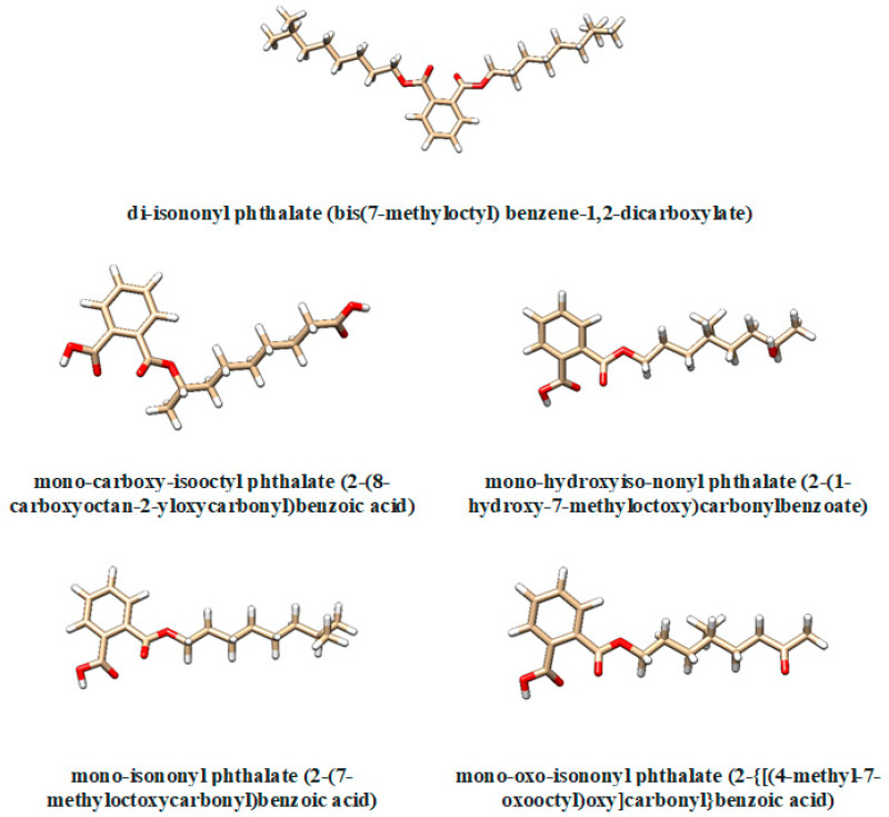
Di-isononyl phthalates are chemicals that are widely used as plasticizers. Humans are extensively exposed to these compounds by dietary intake, through inhalation and skin absorption. Sulfotransferases (SULTs) are enzymes responsible for the detoxification and elimination of numerous endogenous and exogenous molecules from the body. Consequently, SULTs are involved in regulating the biological activity of various hormones and neurotransmitters. The present study considers a computational approach to predict the toxicological potential of the metabolites of di-isononyl phthalate. Furthermore, molecular docking was considered to evaluate the inhibitory potential of these metabolites against the members of family 1 of SULTs. The metabolites of di-isononyl phthalate reveal a potency to cause liver damage and to inhibit receptors activated by peroxisome proliferators. These metabolites are also usually able to inhibit the activity of the members of family 1 of SULTs, except for SULT1A3 and SULT1B1. The outcomes of this study are important for an enhanced understanding of the risk of human exposure to di-isononyl phthalates.
Keywords: di-isononyl phthalate monoesters; metabolism; molecular docking; toxicological effects.
Conflict of interest statement
The authors declare no conflict of interest.
Figures


Similar articles
-
Inhibition of Human Sulfotransferases by Phthalate Monoesters.Front Endocrinol (Lausanne). 2022 Apr 22;13:868105. doi: 10.3389/fendo.2022.868105. eCollection 2022. Front Endocrinol (Lausanne). 2022. PMID: 35528018 Free PMC article.
-
Docking study: PPARs interaction with the selected alternative plasticizers to di(2-ethylhexyl) phthalate.J Enzyme Inhib Med Chem. 2016;31(3):448-55. doi: 10.3109/14756366.2015.1037748. Epub 2015 May 5. J Enzyme Inhib Med Chem. 2016. PMID: 25942360
-
Mono-(3-carboxypropyl) phthalate, a metabolite of di-n-octyl phthalate.J Toxicol Environ Health A. 2006 Feb;69(3-4):215-27. doi: 10.1080/15287390500227381. J Toxicol Environ Health A. 2006. PMID: 16263692
-
NTP Center for the Evaluation of Risks to Human Reproduction: phthalates expert panel report on the reproductive and developmental toxicity of di-isononyl phthalate.Reprod Toxicol. 2002 Sep-Oct;16(5):679-708. doi: 10.1016/s0890-6238(02)00034-5. Reprod Toxicol. 2002. PMID: 12406496 Review. No abstract available.
-
Reproductive toxic potential of phthalate compounds - State of art review.Pharmacol Res. 2021 May;167:105536. doi: 10.1016/j.phrs.2021.105536. Epub 2021 Mar 4. Pharmacol Res. 2021. PMID: 33677105 Review.
Cited by
-
Synthesis, in silico ADMET prediction analysis, and pharmacological evaluation of sulfonamide derivatives tethered with pyrazole or pyridine as anti-diabetic and anti-Alzheimer's agents.Saudi Pharm J. 2024 May;32(5):102025. doi: 10.1016/j.jsps.2024.102025. Epub 2024 Mar 12. Saudi Pharm J. 2024. PMID: 38550332 Free PMC article.
References
-
- Croom E. Metabolism of xenobiotics of human environments. Prog. Mol. Biol. Transl. Sci. 2012;112:31–88. - PubMed
-
- Kurogi K., Rasool M.I., Alherz F.A., El Daibani A.A., Bairam A.F., Abunnaja M.S., Yasuda S., Wilson L.J., Hui Y., Liu M.C. SULT genetic polymorphisms: Physiological, pharmacological and clinical implications. Expert Opin. Drug Metab. Toxicol. 2021;17:767–784. doi: 10.1080/17425255.2021.1940952. - DOI - PMC - PubMed
MeSH terms
Substances
LinkOut - more resources
Full Text Sources
