. 2023 Sep 5;13(18):2501.
doi: 10.3390/nano13182501.

The Potentiating Effect of Graphene Oxide on the Arylhydrocarbon Receptor (AhR)-Cytochrome P4501A (Cyp1A) System Activated by Benzo(k)fluoranthene (BkF) in Rainbow Trout Cell Line

Affiliations

The Potentiating Effect of Graphene Oxide on the Arylhydrocarbon Receptor (AhR)-Cytochrome P4501A (Cyp1A) System Activated by Benzo(k)fluoranthene (BkF) in Rainbow Trout Cell Line

Ana Valdehita et al. Nanomaterials (Basel). .

Abstract

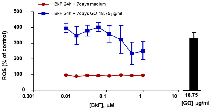
The increasing use of graphene oxide (GO) will result in its release into the environment; therefore, it is essential to determine its final fate and possible metabolism by organisms. The objective of this study was to assess the possible role of the aryl hydrocarbon receptor (AhR)-dependent cytochrome P4501A (Cyp1A) detoxification activities on the catabolism of GO. Our hypothesis is that GO cannot initially interact with the AhR, but that after an initial degradation caused by other mechanisms, small fractions of GO could activate the AhR, inducing Cyp1A. The environmental pollutant benzo(k)fluoranthene (BkF) was used for the initial activation of the AhR in the rainbow trout (Oncorhynchus mykiss) cell line RTL-W1. Pre-, co-, and post-exposure experiments with GO were performed and Cyp1A induction was monitored. The strong stimulation of Cyp1A observed in cells after exposure to GO, when BkF levels were not detected in the system, suggests a direct action of GO. The role of the AhR was confirmed by a blockage of the observed effects in co-treatment experiments with αNF (an AhR antagonist). These results suggest a possible role for the AhR and Cyp1A system in the cellular metabolism of GO and that GO could modulate the toxicity of environmental pollutants.

Keywords: Cyp1A; EROD; aryl hydrocarbon receptor; fish; graphene.


Conflict of interest statement

The authors declare that they have no conflict of interest.

Figures

Figure 1
Figure 1
Effect of graphene oxide (GO) on RTL-W1 cell viability. (A) RTL-W1 cells were exposed to increasing concentrations of GO for 7 days. The cytotoxicity of GO was assessed by means of the AB assay, 5′CFDA-AM assay, and NRU assay. (B) Level of intracellular reactive oxygen species (ROS) upon exposure to increasing concentrations of GO for 7 days. The data points and error bars represent the mean and standard error of the mean (SEM) of at least three independent experiments. Statistically significant differences with respect to the vehicle control (one-way rm ANOVA, Dunnett’s post hoc test) are indicated as follows: * p < 0.05, ** p < 0.01, and *** p < 0.001.
Figure 2
Figure 2
Selected TEM images of RTL-W1 non-exposed cells (A) and 7 days GO exposed at 18.75 µg/mL (BF). (B,C) GO nanoplatelets (black arrows) observed adjacent to the cell surface. (C,D) GO nanoplatelets (black arrows) interacting with the plasma membrane (pm) and penetrating the latter, leading to plasma membrane disruption (the site of disruption is indicated with a white asterisk). (E,F) Intracellular aggregation of GO nanoplatelets (black arrows). Intracellular location of GO throughout the cytoplasm (E) or in membrane-surrounded vesicles (F).
Figure 3
Figure 3
Ethoxyresorufin-O-deethylase (EROD) and Benzyloxy-4-trifluoromethylcoumarin-O-debenzyloxylase (BFCOD) activities after co-exposure (A,B) and pre-exposure (C,D) to GO and BkF. (A,B) RTL-W1 cells were co-exposed to increasing concentrations of Benzo(k)fluoranthene (BkF) (0.001–1 µM) alone or together with 4.6 µg/mL GO or 18.75 µg/mL GO for 7 days. (C,D) cells were pre-exposed to GO at 4.6 µg/mL GO or 18.75 µg/mL GO for 7 days and then to increasing concentrations of BkF (0.00025–0.125 µM) alone for 24 h. The EROD activity levels measured in cell cultures co-exposed and pre-exposed to GO and BkF are shown in (A,C), respectively, and represented as pmol of resorufin/min/mg protein. The BFCOD activity levels measured in cell cultures co-exposed and pre-exposed to GO and BkF are shown in (B,D), respectively, and represented as pmol of HFC/min/mg protein. The data points and error bars represent the mean and standard error of the mean (SEM) of at least three independent experiments.
Figure 4
Figure 4
EROD activity and mRNA expression after post-exposure to GO. (A) EROD activity levels in RTL-W1 cells pre-incubated with increasing concentrations of BkF (0.01–1 µM) for 24 h and post-exposed to media, 4.6 µg/mL, or 18.75 µg/mL of GO for 7 days. The EROD activity levels are represented as pmol/min/mg protein. cyp1A (B) and ahr (C) mRNA expression levels in RTL-W1 cells pre-incubated with 0.01, 0.25, and 1 µM of BkF for 24 h and post-exposed to media or to 18.75 mg/mL GO for 7 days. cyp1A and ahr mRNA expression are represented as a fold expression with respect to the corresponding mRNA expression level in control cells. Statistically significant differences with respect to the control cells (vehicle) are indicated as * for p < 0.05 and *** for p < 0.001 and with respect to GO 7 days as + for p < 0.05 and +++ for p < 0.001. Statistically significant differences between mRNA cyp1A levels measured for the same BkF concentration with and without post-exposure to GO 18.75 mg/mL for 7 days are indicated as # and ### (p < 0.05 and <0.001) (One-way rm ANOVA, Bonferroni’s Multiple Comparison test). Data points and error bars represent the mean and standard error of the mean (SEM) of at least three independent experiments.
Figure 5
Figure 5
cyp1A (A) and ahr (B) mRNA expression levels. RTL-W1 cells were pre-incubated for 1 h with 1 µM of α-Naphtoflavone (αNF), then exposed to BkF (0.25 µM) for 24 h more, and finally post-exposed to GO 18.75 µg/mL for 7 days. mRNA expression levels are represented as a fold expression with respect to the levels measured in the control cells. Statistically significant differences with respect to the vehicle control are indicated as: * for p < 0.05 and *** for p < 0.001; with respect to GO 7 days as # for p < 0.05 and ### for p < 0.001; with respect to BkF 0.25 µM for 24 h then 7 days with media as αα for p < 0.01; and with respect to BkF 0.25 µM for 24 h then 7 days with GO 18.75 mg/mL as ++ for p < 0.01 and +++ for p < 0.001 (One-way rm ANOVA, Bonferroni’s Multiple Comparison test). Bars and error bars represent the mean and standard error of the mean (SEM) of three independent experiments.
Figure 6
Figure 6
Levels of intracellular reactive oxygen species (ROS) upon post-exposure conditions to GO. RTL-W1 cells were pre-incubated with increasing concentrations of BkF (0.078–1 µM) for 24 h and post-exposed 7 days with media or with GO at 18.75 µg/mL. The bar represents the value of ROS after exposure to GO at 18.75 µg/mL alone. Point, bar, and errors represent the mean and standard error of the mean (SEM) of three independent experiments.

Similar articles

Cited by

References

    1. Novoselov K.S., Geim A.K., Morozov S.V., Jiang D., Zhang Y., Dubonos S.V., Grigorieva I.V., Firsov A.A. Electric field effect in atomically thin carbon films. Science. 2004;306:666–669. doi: 10.1126/science.1102896. - DOI - PubMed
    1. Halim A., Luo Q., Ju Y., Song G. A mini review focused on the recent applications of graphene oxide in stem cell growth and differentiation. Nanomaterials. 2018;8:736. doi: 10.3390/nano8090736. - DOI - PMC - PubMed
    1. Singh D.P., Herrera C.E., Singh B., Singh S., Singh R.K., Kumar R. Graphene oxide: An efficient material and recent approach for biotechnological and biomedical applications. Mater. Sci. Eng. C Mater. Biol. Appl. 2018;86:173–197. doi: 10.1016/j.msec.2018.01.004. - DOI - PubMed
    1. Joshi K., Mazumder B., Chattopadhya P., Bora N.S., Goyary D., Karmakar S. Graphene family of nanomaterials: Reviewing advanced applications in drug delivery and medicine. Curr. Drug Deliv. 2019;16:195–214. - PubMed
    1. Chen L., Li J., Chen Z., Gu Z., Yan L., Zhao F., Zhang A. Toxicological Evaluation of Graphene-Family Nanomaterials. J. Nanosci. Nanotechnol. 2020;20:1993–2006. doi: 10.1166/jnn.2020.17364. - DOI - PubMed

LinkOut - more resources