. 2023 Sep 12;13(18):2546.
doi: 10.3390/nano13182546.

The Apoptosis Inhibitor Protein Survivin Is a Critical Cytoprotective Resistor against Silica-Based Nanotoxicity

Affiliations

The Apoptosis Inhibitor Protein Survivin Is a Critical Cytoprotective Resistor against Silica-Based Nanotoxicity

Christina Breder-Bonk et al. Nanomaterials (Basel). .

Abstract

Exposure to nanoparticles is inevitable as they become widely used in industry, cosmetics, and foods. However, knowledge of their (patho)physiological effects on biological entry routes of the human body and their underlying molecular mechanisms is still fragmented. Here, we examined the molecular effects of amorphous silica nanoparticles (aSiNPs) on cell lines mimicking the alveolar-capillary barrier of the lung. After state-of-the-art characterization of the used aSiNPs and the cell model, we performed cell viability-based assays and a protein analysis to determine the aSiNP-induced cell toxicity and underlying signaling mechanisms. We revealed that aSiNPs induce apoptosis in a dose-, time-, and size-dependent manner. aSiNP-induced toxicity involves the inhibition of pro-survival pathways, such as PI3K/AKT and ERK signaling, correlating with reduced expression of the anti-apoptotic protein Survivin on the protein and transcriptional levels. Furthermore, induced Survivin overexpression mediated resistance against aSiNP-toxicity. Thus, we present the first experimental evidence suggesting Survivin as a critical cytoprotective resistor against silica-based nanotoxicity, which may also play a role in responses to other NPs. Although Survivin's relevance as a biomarker for nanotoxicity needs to be demonstrated in vivo, our data give general impetus to investigate the pharmacological modulation of Survivin`s functions to attenuate the harmful effects of acute or chronic inhalative NP exposure.

Keywords: alveolar-capillary barrier; amorphous silica nanoparticles; cytotoxic response; inflammation; lung model; nanotoxicity.


Conflict of interest statement

The authors declare no conflict of interest.

Figures

Figure 1
Figure 1
Characterization of the used amorphous silica NP system (aSiNP) by (Cryo-)transmission electron microscopy (TEM). Representative pictures of analyzed aSiNPs are shown in overview (upper row) and higher magnification (lower row): (A) aSiNP15 (TEM), (B) aSiNP15 (Cryo-TEM), (C) aSiNP L (TEM), and (D) aSiNP125 (TEM). Scale bars, 100 nm.
Figure 2
Figure 2
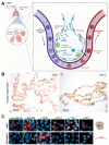
Biological model to mimic the alveolar-capillary barrier of the lung: (A) Illustration of a bronchial strain as the first protective cellular barrier against inhaled nanoparticle-containing air. (B) Light microscopic analysis of biomarker expression. Expression of CD31 and TTF-1 was analyzed in epithelial human lung tissue by immunohistochemistry with specific antibodies (brown). HE (hematoxylin/eosin) staining was applied to visualize tissue morphology (blue). CD31 surface marker was present on the membrane of epithelial cells in the human lung tissue, whereas TTF-1 was prominently expressed in cell nuclei. (C) Marker profiling of the H441 and ISO-HAS-1 dual cell line model was carried out by fluorescence microscopy after staining with specific antibodies (α-E-cadherin, α-EpCAM, α-CD31, α-SP-A, α-TTF-1, α-ZO-1) and the nuclear stain Hoechst-33342 (blue) to evaluate the cellular differentiation state. Scale bars, 10 µm. Respective cell types are symbolized by referring to (A). Created with BioRender.com.
Figure 3
Figure 3
Interaction and internalization of aSiNPs by epithelial lung cells: (A) Immunofluorescence and light microscopy of H441 cells after incubation with increasing concentrations of fluorescent aSiNP (Kisker red) for 2 h in serum-deprived medium. Scale bars, 10 µm. (BD) aSiNP15 nanoparticle uptake by H441 cells was visualized by TEM. H441 cells were treated with 1012 NP/mL for 2 h in serum-containing (B) or serum-free medium (C,D). Representative images are shown in overview (upper row), and in detail view (lower row) indicating the presence (B) or absence (C,D) of a protein corona around aSiNP15. Endocytic vesicles are marked by arrows. Scale bars, 500 nm (upper panel), and 100 nm (lower panel).
Figure 4
Figure 4
aSiNP15 treatment induced dose- and time-dependent toxicity on a dual alveolar-capillary barrier model. ISO-HAS-1 and H441 cells were treated with different particle concentrations in serum-free medium for 4 h; for later time points, treated cells were cultured for additional 20 h in serum-containing medium: (A) Live and dead staining analyzed by fluorescence microscopy. Green fluorescence marks viable cells; red fluorescence visualizes dead cells. (B) CellTiter-Glo™ Viability Assay. Data were normalized to untreated control samples for each time point. Unpaired t-test was used to compare treated cells to untreated control groups. Column, mean; bars, ±S.D. ***, p < 0.001, ****, p < 0.0001. (C,D) aSiNP15 induced apoptosis in ISO-HAS-1 and H441 cells via activation of caspase-3. (C) Light microscopic analysis of apoptosis marker caspase-3. Immunohistochemical staining of ISO-HAS-1 (upper panel) and H441 (lower panel) revealed expression of cleaved caspase-3 as a marker for apoptosis after 4 h of aSiNP15 treatment. Briefly, cells were treated with 1012 NP/mL for 4 or 24 h, fixed, stained for cleaved α-caspase-3, and visualized by light microscopy. Scale bars, 150 µm. (D) Quantification of in vitro caspase-3 activity confirmed time- and dose-dependent activation of apoptosis. Cleaved α-caspase-3 assay was performed with untreated and aSiNP15-treated cells. Cells were incubated for 4 h and 24 h with 1010 or 1012 NP/mL as indicated. Data were normalized to untreated control samples for each time point. Unpaired t-test was used to compare treated cells to untreated control groups. Column, mean; bars, ±S.D. *, p < 0.05, **, p < 0.01, ****, p < 0.0001.
Figure 5
Figure 5
High concentrations of aSiNP15 attenuate PI3K/Akt and ERK signaling. (A,B) Activation of PI3K/Akt and ERK signaling pathways was analyzed by assessing the level of phosphorylated PI3K/Akt (pAkt) and ERK (pERK; Thr202/Tyr204) by immunoblot analysis. Whereas sub-toxic NP concentrations (1010 NP/mL) had no effect (B), pAkt and pERK decreased after incubation with 1012 NP/mL. ISO-HAS-1 and H441 were treated with 1012 NP/mL (A) or 1010 NP/mL (B) aSiNP15 (indicated by “+”), or in serum-deprived medium as control (indicated by “−“) for 2 h, 4 h, and 6 h. Proteins were detected by (phosphorylation-) specific antibodies, the respective non-phosphorylated proteins, and Actin served as loading controls. (C) Relevance of ERK signaling was confirmed by combined MEK (mitogen-activated protein kinase kinase) inhibitor treatment. After H441 cells were pre-treated for 2 h with or without the MEK inhibitor UO126 (10 µM), treatment with 1010, 1011, or 1012 NP/mL of type aSiNP15 was conducted for 4 h, 8 h and 24 h. Cell viability was measured via an MTT assay. Data were normalized for each time point to untreated controls. (*) Unpaired t-test was performed to compare MEK inhibitor-treated samples to samples with similar treatment without inhibitor. Columns, mean; bars, ±S.D. *, p < 0.05, **, p < 0.01, ***, p < 0.001, ns, not significant.
Figure 6
Figure 6
aSiNP-mediated toxicity was accompanied by dose-dependent downregulation of the anti-apoptotic protein Survivin: (AF) Immunoblot analysis revealed dose-dependent downregulation of Survivin in response to treatment with aSiNP15. ISO-HAS-1 and H441 cells were treated with (“+”) 1012 NP/mL (AC) or 1010 NP/mL (DF) aSiNP15 or without particles (“−“) for 0 and 2 h in serum-free medium. For incubation times longer than 4 h, serum-containing medium was added after 4 h to prevent starvation-induced cell death. Proteins were detected by specific antibodies. β-Actin served as loading control. (G) Suppression of the Survivin promoter was revealed by a luciferase reporter assay following aSiNP15 treatment. Briefly, H441 cells were treated with 1010, 1011, and 1012 NP/mL for 4 h in serum-free medium. Likewise, for the 24 h time point, serum-containing medium was added after 4 h. After incubation, cells were lysed, and lysates were used for quantification of luciferase activity as described [36]. Data were normalized to untreated controls, and unpaired t-test was performed. Columns, mean; bars, ± S.D. *, p < 0.05, ****, p < 0.0001.
Figure 7
Figure 7
Overexpression of Survivin counteracts aSiNP15-induced cell death: (A) In stably Survivin-GFP-expressing H441 cells (H441 Surv), Survivin exhibits its characteristic localization during mitotic phases shown via immunofluorescence microscopy (as indicated). In prophase/metaphase, it localizes at chromatin sides. In anaphase, Survivin dissociates from chromatin and associates with the midzone spindle apparatus. In telophase, it localizes at the midbody, and disperses in the cytoplasm and/or in the nucleus during interphase. Scale bars, 10µm. (B) Morphological stability of H441 cells after treatment with different aSiNP15 concentrations at the beginning of treatment (0 h) and after 4 h aSiNP15 exposure. Cell morphology was visualized by light microscopy. Scale bars, 150 µm. (C) High doses of aSiNP15 induce cell death after long-term incubation. An MTT viability assay of stably expressing Survivin–GFP H441 cells was performed after 4 h, 8 h, and 24 h of aSiNP15 treatment as indicated. Data were normalized to untreated controls, and unpaired t-test was performed. Columns, mean; bars, ±S.D. ***, p < 0.001, ****, p < 0.0001. (D) CellTiter-GloTM viability assay of H441 Surv (grey) and control cells (H441, black) was performed after 4 h, 8 h, and 24 h of aSiNP15 treatment as indicated. Data were normalized to untreated controls, and unpaired t-test was performed. Columns, mean; bars, ±S.D. **, p < 0.01, ***, p < 0.001.
Figure 8
Figure 8
Survivin is physiologically expressed in epithelial cells of the human and mouse lung. Immunohistochemical staining of Survivin (A,C) and TTF-1 (B,D) in epithelial human and mouse lung tissue with specific antibodies (brown) and visualized by light microscopy. HE staining was applied to visualize lung tissue structure (blue). Survivin localized in the nuclei and cytoplasm of mouse and human epithelial lung cells (A,C), whereas TTF-1 was prominently overexpressed in the nuclei (B,D). Scale bars, 50 µM.

Similar articles

Cited by

References

    1. Davies J.C.T. PEN 13-Nanotechnology Oversight. [(accessed on 14 January 2019)]. Available online: https://www.wilsoncenter.org/publication/pen-13-nanotechnology-oversight.
    1. Liljenström C.L.D., Finnveden G. Silicon-Based Nanomaterials in a Life-Cycle Perspective, Including a Case Study on Self-Cleaning Coatings. 2013. [(accessed on 14 January 2019)]. Available online: https://www.researchgate.net/publication/280264076_Silicon-based_nanomat....
    1. Vance M., Lawson N.C., Rupal M., Beck P., Burgess J.O. Color and Gloss of Nano-Filled Resin-Modified Glass Ionomers and Resin Composites. J. Esthet. Restor. Dent. 2015;27:293–299. doi: 10.1111/jerd.12118. - DOI - PubMed
    1. Croissant J.G., Fatieiev Y., Khashab N.M. Degradability and Clearance of Silicon, Organosilica, Silsesquioxane, Silica Mixed Oxide, and Mesoporous Silica Nanoparticles. Adv. Mater. 2017;29:1604634. doi: 10.1002/adma.201604634. - DOI - PubMed
    1. Dekkers S., Krystek P., Peters R.J., Lankveld D.P., Bokkers B.G., van Hoeven-Arentzen P.H., Bouwmeester H., Oomen A.G. Presence and risks of nanosilica in food products. Nanotoxicology. 2011;5:393–405. doi: 10.3109/17435390.2010.519836. - DOI - PubMed

LinkOut - more resources