. 2023 Sep 4;12(9):1128.
doi: 10.3390/pathogens12091128.

Modulation of Macrophage Redox and Apoptotic Processes to Leishmania infantum during Coinfection with the Tick-Borne Bacteria Borrelia burgdorferi

Affiliations

Modulation of Macrophage Redox and Apoptotic Processes to Leishmania infantum during Coinfection with the Tick-Borne Bacteria Borrelia burgdorferi

Danielle Pessôa-Pereira et al. Pathogens. .

Abstract

Canine leishmaniosis (CanL) is a zoonotic disease caused by protozoan Leishmania infantum. Dogs with CanL are often coinfected with tick-borne bacterial pathogens, including Borrelia burgdorferi in the United States. These coinfections have been causally associated with hastened disease progression and mortality. However, the specific cellular mechanisms of how coinfections affect microbicidal responses against L. infantum are unknown. We hypothesized that B. burgdorferi coinfection impacts host macrophage effector functions, prompting L. infantum intracellular survival. In vitro experiments demonstrated that exposure to B. burgdorferi spirochetes significantly increased L. infantum parasite burden and pro-inflammatory responses in DH82 canine macrophage cells. Induction of cell death and generation of mitochondrial ROS were significantly decreased in coinfected DH82 cells compared to uninfected and L. infantum-infected cells. Ex vivo stimulation of PBMCs from L. infantum-seronegative and -seropositive subclinical dogs with spirochetes and/or total Leishmania antigens promoted limited induction of IFNγ. Coexposure significantly induced expression of pro-inflammatory cytokines and chemokines associated with Th17 differentiation and neutrophilic and monocytic recruitment in PBMCs from L. infantum-seropositive dogs. Excessive pro-inflammatory responses have previously been shown to cause CanL pathology. This work supports effective tick prevention and risk management of coinfections as critical strategies to prevent and control L. infantum progression in dogs.

Keywords: Lyme disease; apoptosis; canine leishmaniosis; coinfection; inflammation; progression.


Conflict of interest statement

The authors declare no conflict of interest.

Figures

Figure 1
Figure 1
B. burgdorferi coinfection increases L. infantum infection rates and cellular burden. DH82 cells were exposed to L. infantum (1:10 MOI) for 24 h, and then exposed to live B. burgdorferi (1:25 MOI) for indicated time points. Cells were fixed with methanol and stained using HEMA 3 solutions. Frequencies of L. infantum-infected cells (A) and number of amastigotes (B) per 100 DH82 post-coinfections with live B. burgdorferi were assessed through light microscopy (100×). Blind and independent assessments were performed by two researchers. Data represent mean ± SEM of three independent experiments, performed in triplicates for each condition. N = 10–23 coverslips per group. Two-way ANOVA, Tukey’s multiple comparisons test. p **** < 0.0001. Li: L. infantum infection. Li + Bb: L. infantum and B. burgdorferi coinfection.
Figure 2
Figure 2
B. burgdorferi exposure decreases late apoptosis/necrosis rates in L. infantum-infected cells. Apoptosis assay using flow cytometry analysis of Annexin V and propidium iodide (PI) staining. DH82 cells were labeled with Annexin V (A488) and 1 µM PI at 24 h after no treatment; L. infantum infection (1:10 MOI); B. burgdorferi infection (1:25 MOI); coinfection, and 10 µM camptothecin (CPT; positive control). (A) Representative scatter plots of Annexin V (x-axis) vs. PI (y-axis). Bottom left quadrant: viable cells (Annexin V−/PI−); bottom right (Annexin V+/PI−): cells undergoing early apoptosis; top right (Annexin V+/PI+): cells that are in late-stage apoptosis/necrosis; top left (Annexin V−/PI+): dead cells. (B) Frequencies of DH82 cells undergoing cell death after infections and treatments. Ordinary one-way ANOVA, Tukey’s multiple comparisons test. p * = 0.0402 (Li vs. Bb); 0.0359 (Li vs. Li + Bb). p **** < 0.0001. (C) Frequencies of early apoptotic and late apoptotic/necrotic DH82 cells after infections and treatments. Two-way ANOVA, Sidak’s multiple comparisons test. Data are presented as mean ± SEM of triplicate experiments. p * = 0.01; p **** < 0.0001. Unstim: unstimulated (media); Li: L. infantum infection; Bb: B. burgdorferi infection; Li + Bb: L. infantum and B. burgdorferi coinfection. Apop.: apoptosis.
Figure 3
Figure 3
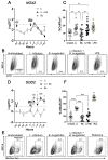
Altered production of reactive oxygen species (ROS) during L. infantum and B. burgdorferi coinfection in DH82 cells. Cells were exposed to L. infantum promastigotes and/or B. burgdorferi spirochetes. Gene expression of NOX2 (A) and SOD2 (D) were quantified via quantitative PCR and normalized using ACTB as an endogenous control. Data represent log transformed-fold change mean values relative to uninfected DH82 cells ± SEM of three independent experiments, with significance assessed via two-way ANOVA, Tukey’s multiple comparisons test. * L. infantum and B. burgdorferi coinfections vs. L. infantum infection. +B. burgdorferi infection vs. L. infantum infection. (A) p ** = 0.002, p ++ = 0.0014, (D) p * = 0.0114, p + = 0.0157, p ++ = 0.0022; p *** = 0.0006, p +++ = 0.0002, p ****/++++ < 0.0001. Representative scatter plots showing percentages of CellROXhi (B) and MitoSOXhi (E) populations after exposure to medium, L. infantum promastigotes, B. burgdorferi spirochetes, and respective positive controls (LPS and rotenone). Percentage of CellROXhi (C) and MitoSOXhi (F) cells after B. burgdorferi exposure and indicated stimulations. Statistical significance was estimated using ordinary one-way ANOVA with post hoc Tukey test. (C) p ** = 0.0071 (unstimulated vs. Li/Li + Bb), 0.0016 (Bb vs. Li+Bb), (F) p **** < 0.0001, mean ± SEM of triplicate experiments.
Figure 4
Figure 4
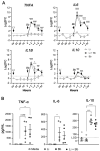
B. burgdorferi induces pro- and anti-inflammatory cytokines gene expression and production in L. infantum-infected DH82 cells. (A) Gene expression of TNFA, IL6, IL1B, and IL10 were quantified via qPCR and normalized using ACTB as an endogenous control. Data represent log transformed-fold change mean values relative to uninfected DH82 cells ± SEM of three independent experiments using Two-way ANOVA, Tukey’s multiple comparisons test. * L. infantum and B. burgdorferi coinfection vs. L. infantum infection. +B. burgdorferi infection vs. L. infantum infection. TNFA, p +++ = 0.0005; p ****/++++ < 0.0001; IL6, p ****/++++ < 0.0001; IL1B, p * = 0.0126; p *** = 0.0003; p +++ = 0.0002; p ****/++++ < 0.0001); IL10, p *** = 0.0007 (2 h), 0.0002 (4 h); p +++ = 0.0008; p ++++ < 0.0001). (B) Supernatants from DH82 cells were collected 24 h after exposure to B. burgdorferi. Release of TNF-α, IL-6, and IL-10 were quantified using Milliplex ELISA. Data represent mean values ± SEM of five independent experiments. Ordinary one-way ANOVA with Tukey post hoc test. TNF-α, p * = 0.0357; IL-6, p * = 0.0394; IL-10, p **** < 0.0001.
Figure 5
Figure 5
B. burgdorferi modulates gene expression and production of pro-inflammatory and regulatory cytokines in PBMCs from L. infantum-seronegative and seropositive dogs. PBMCs were stimulated with live B. burgdorferi spirochetes (1:10 MOI) for different timepoints. Cells were harvested at 24 h after infections for gene expression analyses. Supernatants were collected at 72 h after infections for ELISA. (A) Gene expression TNFA, IL6, IL1B, IL12A, IL12B, IFNG, and IL10 were quantified by quantitative PCR and normalized using ACTB as an endogenous control. Data represent log transformed-fold change mean values (relative to unstimulated PBMCs from L. infantum-seronegative dogs) ±SEM of three independent experiments. Kruskal–Wallis with Dunn’s post hoc test. TNFA, p ** = 0.0027; IL6, p * = 0.0354; p ** = 0.005; IL1B, p ** = 0.0015; IL12B, p * < 0.05; IL10, p ** = 0.0089. N = 5–6 samples per group. (B) Release of TNF-α, IL-6, IFNγ, and IL-10 were quantified using Milliplex ELISA. Data represent mean values ± SEM of five independent experiments. Wilcoxon matched-pairs rank test, Holm–Sidak method. Adjusted p * = 0.03125. N = 5–8 samples per group.
Figure 6
Figure 6
B. burgdorferi modulates gene expression and production of Type 17-related cytokines/chemokines gene expression in PBMCs from L. infantum-seronegative and seropositive dogs. (A) Gene expression of IL23p19, TGFB, IL17A, and IL22 were quantified by quantitative PCR and normalized using ACTB as an endogenous control. Data represent log transformed-fold change mean values relative to unstimulated PBMCs from L. infantum-seronegative dogs ± SEM of three independent experiments. Kruskal–Wallis with Dunn’s post hoc test. IL12p19, p ** = 0.0027; IL17A/IL22, p * = 0.0199; p ** = 0.0089. (B) Release of IL-17A via sandwich ELISA, IL-8, CXCL1, and CCL2 via Milliplex ELISA. Data represent mean values ± SEM of five independent experiments. Wilcoxon matched-pairs rank test, Holm-Sidak method. Adjusted p * < 0.03125.

Similar articles

Cited by

References

    1. Burza S., Croft S.L., Boelaert M. Leishmaniasis. Lancet. 2018;392:951–970. doi: 10.1016/S0140-6736(18)31204-2. - DOI - PubMed
    1. Bogdan C. Macrophages as host, effector and immunoregulatory cells in leishmaniasis: Impact of tissue micro-environment and metabolism. Cytokine X. 2020;2:100041. doi: 10.1016/j.cytox.2020.100041. - DOI - PMC - PubMed
    1. Kumar R., Nylén S. Immunobiology of visceral leishmaniasis. Front. Immunol. 2012;3:251. doi: 10.3389/fimmu.2012.00251. - DOI - PMC - PubMed
    1. Gavgani A.S.M., Hodjati M., Mohite H., Davies C. Effect of insecticide-impregnated dog collars on incidence of zoonotic visceral leishmaniasis in Iranian children: A matchedcluster randomised trial. Lancet Lond. Engl. 2002;360:374–379. doi: 10.1016/S0140-6736(02)09609-5. - DOI - PubMed
    1. Dantas-Torres F. The role of dogs as reservoirs of Leishmania parasites, with emphasis on Leishmania (Leishmania) infantum and Leishmania (Viannia) braziliensis. Vet. Parasitol. 2007;149:139–146. doi: 10.1016/j.vetpar.2007.07.007. - DOI - PubMed

LinkOut - more resources