. 2023 Sep 6;12(9):1141.
doi: 10.3390/pathogens12091141.

A Multilayered Imaging and Microfluidics Approach for Evaluating the Effect of Fibrinolysis in Staphylococcus aureus Biofilm Formation

Affiliations

A Multilayered Imaging and Microfluidics Approach for Evaluating the Effect of Fibrinolysis in Staphylococcus aureus Biofilm Formation

Raffaella Parente et al. Pathogens. .

Abstract

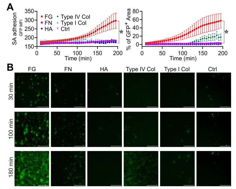
The recognition of microbe and extracellular matrix (ECM) is a recurring theme in the humoral innate immune system. Fluid-phase molecules of innate immunity share regulatory roles in ECM. On the other hand, ECM elements have immunological functions. Innate immunity is evolutionary and functionally connected to hemostasis. Staphylococcus aureus (S. aureus) is a major cause of hospital-associated bloodstream infections and the most common cause of several life-threatening conditions such as endocarditis and sepsis through its ability to manipulate hemostasis. Biofilm-related infection and sepsis represent a medical need due to the lack of treatments and the high resistance to antibiotics. We designed a method combining imaging and microfluidics to dissect the role of elements of the ECM and hemostasis in triggering S. aureus biofilm by highlighting an essential role of fibrinogen (FG) in adhesion and formation. Furthermore, we ascertained an important role of the fluid-phase activation of fibrinolysis in inhibiting biofilm of S. aureus and facilitating an antibody-mediated response aimed at pathogen killing. The results define FG as an essential element of hemostasis in the S. aureus biofilm formation and a role of fibrinolysis in its inhibition, while promoting an antibody-mediated response. Understanding host molecular mechanisms influencing biofilm formation and degradation is instrumental for the development of new combined therapeutic approaches to prevent the risk of S. aureus biofilm-associated diseases.

Keywords: Staphylococcus aureus; biofilm; extracellular matrix; fibrinolysis; hemostasis; innate immunity.


Conflict of interest statement

The authors declare no conflict of interest.

Figures

Figure 1
Figure 1
Role of fibrinogen in S. aureus (SA) adhesion. (A,B) The bottom surface of microfluidic channels coated with different ECM molecules was used. Fibrinogen (FG, 100 µg/mL), Fibronectin (FN, 100 µg/mL), Hyaluronic acid (HA, 100 µg/mL), type I collagen (100 µg/mL) and type IV collagen (100 µg/mL). Flux (0.5 µL/min) of S. aureus (1.7 × 10⁶ CFU/mL) in TSB. (A) GFP MFI ± SE and Mean % of GFP+ area ± SE overtime. Each point refers to the average of 3,6 ROIs (regions of interest) from n = 2 experiments performed. * p < 0.05 FG vs. ctrl (Dunn’s Test). (B) GFP images are shown at representative time points (t = 30, 100 and 180 min) referring to one experiment. Bar, 100 µm.
Figure 2
Figure 2
Role of fibrinogen in the assembly and formation of S. aureus (SA) biofilm. (AD) S. aureus (1.7 × 10⁷ CFU/mL) previously adhered on the bottom surface of straight microfluidic channels. Conditions include TSB, 10% of normal (NP) and fibrinogen-depleted (FGˉ) ACD-plasma diluted in TSB and FGˉACD-plasma diluted in TSB added with human purified FG (400 µg/mL) and FG (400 µg/mL) in TSB. (A) S. aureus growth and biofilm formation were evaluated, respectively, as relative GFP (upper) and PI (lower) MFI. Values are represented as functions of time and normalized over the first time point. The PI signal was considered superimposed to the GFP-positive mask, as described in Materials and Methods. * p < 0.05, ** p < 10−3 FGˉ vs. NP at t = 180 min, n = 12, 15, Dunn’s test. (B) Images of bright field (BF), GFP and PI at representative time points (t = 30, 100 and 180 min), referring to one experiment. At t = 100 min, BF close-up images representing S. aureus morphology are also shown. Bar, 50 µm. (C) Mean CI of S. aureus colonies *** p < 10−4 FGˉ vs. NP at t = 100 min. (D) Relative GFP MFI over time after the flux increase (at t = 200 min, 10 µL/min for 30 min to 50 µL/min until the end of the experiment). *** p < 10−4 FGˉ vs. NP, n = 9, 12, Dunn’s test. (A,C) Mean ± SE of 9 to 15 ROIs from three experiments out of four performed with similar results. (D) Mean ± SE of 3 to 12 ROIs from two experiments out of four performed with similar results.
Figure 3
Figure 3
Plasminogen does not affect S. aureus (SA) biofilm formation. (AC) Same experimental setting and analysis as those in Figure 2 were used; 10% of PLG-depleted (PLGˉ) or FGˉ and NP ACD-plasma diluted in TSB were used. (A) Relative GFP (upper) and PI (lower) MFI ± SE over time. * p < 0.05 PLGˉ vs. FGˉ, t = 180 min, Dunn’s test. (B) BF, GFP and PI images at t = 30, 100 and 180 min, representative of one experiment. Bar, 50 µm. (C) Mean CI of S. aureus colonies. (A,C) Each point refers to the Mean ± SE of three ROIs from one experiment of three performed.
Figure 4
Figure 4
Role of fibrinogen in the adhesion, assembly and formation of biofilm by S. aureus (SA) in flow. (AC) A pillar-based microfluidic device was used. S. aureus (1.7 × 10⁶ CFU/mL) was injected with a flow rate of 0.5 µL/min at the same experimental conditions as in Figure 2. (A) Representative BF images which refer to one experiment of six performed, showing different initiation phases (adhesion, coagulation, fibrin matrix assembly) that lead to biofilm formation around the micropillar. (B) S. aureus growth as GFP MFI (×103) and biofilm detection as PI MFI (×103) as a function of time. Each point refers to the Mean ± SE of 6–15 ROIs from three experiments. Missing values in FGˉ correspond to time points where less than two pillars (over six) had a detectable signal, as described in the Section 2. * p < 0.05 FGˉ + FG vs. FGˉ at t = 180 min, Dunn’s test. (C) GFP, PI and contrast images are shown at t = 30, 100 and 180 min. Bar, 50 µm. (A,C) Arrow indicates the flow direction.
Figure 5
Figure 5
Triggering fibrinolysis interferes in the formation of S. aureus (SA) biofilm. (AD) Straight microfluidic channels, with S. aureus (1.7 × 10⁷ CFU/mL) previously adhered on the bottom surface, were used in the experiments (n = 2); 10% of NP with or without the addition of recombinant purified uPA (0.4 µg/mL) or tPA (0.4 µg/mL) and FGˉ were used. (A) S. aureus growth as relative GFP MFI ± SE (upper) and biofilm formation as PI MFI ± SE (lower) over the initial time. (B) BF, GFP and images referring to one representative experiment are shown at t = 30, 100 and 180 min. Bar, 50 µm. (C) CI of S. aureus colonies expressed as the Mean ± SE. (D) S. aureus detachment as an expression of the relative GFP after the flow rate was increased up to 10 µL/min for 30 min and later to 50 µL/min until the end of the acquisition. Each point is the Mean ± SEM of three to eight ROIs from one experiment out of two performed with similar results. * p < 0.05 NP vs. NP + uPA at t = 180 min (A), t > 100 min (C), t = 240 min (D), Dunn’s test.
Figure 6
Figure 6
Triggering fibrinolysis interferes in the initial phase, leading to biofilm formation by S. aureus (SA) in flow. (A,B) A pillar-based microfluidic device and the same experimental conditions as those in Figure 4 were used; 10% of NP with or without the addition of recombinant purified uPA (0.4 µg/mL) or tPA (0.4 µg/mL) and FGˉ were used. (A) S. aureus growth (left) as GFP MFI (×103) and biofilm detection (right) as PI MFI (×103) as a function of time. Each point refers to the Mean ± SE of 8–10 ROIs from two experiments. * p < 0.05, ** p < 0.005 NP vs. NP + tPA, n = 10, t = 220 min. (B) GFP, PI and contrast images (t = 30, 100 and 180 min) of one representative experiment. Bar 50 µm. The arrow indicates the flow direction.
Figure 7
Figure 7
Reactivation of fibrinolysis in S. aureus-induced sepsis favors IgG-mediated pathogen killing. (AC) Microfluidic channels and S. aureus (SA) (1.7 × 10⁷ CFU/mL) that were previously adhered were used; 10% of ACD-plasma of PzP 1 (higher titer of IgG anti-SA) (A) and PzP 2 (lower titer of IgG anti-SA) (B) in combination with uPA (0.4 µg/mL) or tPA (0.4 µg/mL) diluted in TSB were fluxed (0.5 µL/min). PzP 1 and PzP 2 ACD-plasma depleted of IgGs (IgGˉ) was also used to ascertain an anti-S. aureus IgG-mediated pathogen killing. (A,B) Upper: SA growth plotted as relative GFP MFI ± SE as a function of time. (A,B) Lower: S. aureus killing as PI MFI ± SE, considered superimposed to a GFP-positive mask, as described in the Section 2. A log scale was used to improve data visualization. (A,B) Each point refers to three to eight ROIs from one experiment (PzP 1) and three to eight ROIs from one experiment (PzP 2). (A,B) * p < 0.05 PzP 2 vs. PzP 2 IgGˉ (n = 3, 4); ** p < 0.01 PzP 1 vs. PzP 1 IgGˉ (n = 4, 8), at t = 220 min, Dunn’s test. (C) GFP and PI images of an experiment involving PzP 1 at t = 30, 100 and 180 min. Bar, 50 µm.

References

    1. Bergmann S., Hammerschmidt S. Fibrinolysis and host response in bacterial infections. Thromb. Haemost. 2007;98:512–520. doi: 10.1160/TH07-02-0117. - DOI - PubMed
    1. Levi M., Van Der Poll T., Büller H.R. Bidirectional relation between inflammation and coagulation. Circulation. 2004;109:2698–2704. doi: 10.1161/01.CIR.0000131660.51520.9A. - DOI - PubMed
    1. Koyama K., Madoiwa S., Nunomiya S., Koinuma T., Wada M., Sakata A., Ohmori T., Mimuro J., Sakata Y. Combination of thrombin-antithrombin complex, plasminogen activator inhibitor-1, and protein C activity for early identification of severe coagulopathy in initial phase of sepsis: A prospective observational study. Crit. Care. 2014;18:R13. doi: 10.1186/cc13190. - DOI - PMC - PubMed
    1. Milbrandt E.B., GenIMS Investigators. Reade M.C., Lee M., Shook S.L., Angus D.C., Kong L., Carter M., Yealy D.M., Kellum J.A. Prevalence and significance of coagulation abnormalities in community-acquired pneumonia. Mol. Med. 2009;15:438–445. doi: 10.2119/molmed.2009.00091. - DOI - PMC - PubMed
    1. Bokarewa M.I., Tarkowski A. Thrombin generation and mortality during Staphylococcus aureus sepsis. Microb. Pathog. 2001;30:247–252. doi: 10.1006/mpat.2000.0425. - DOI - PubMed

LinkOut - more resources