. 2023 Sep 15;16(9):1306.
doi: 10.3390/ph16091306.

Neuroprotective Effect of Nosustrophine in a 3xTg Mouse Model of Alzheimer's Disease

Affiliations

Neuroprotective Effect of Nosustrophine in a 3xTg Mouse Model of Alzheimer's Disease

Iván Carrera et al. Pharmaceuticals (Basel). .

Abstract

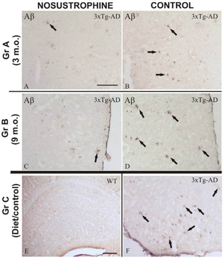
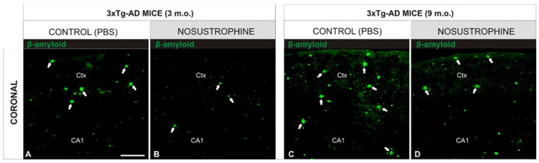
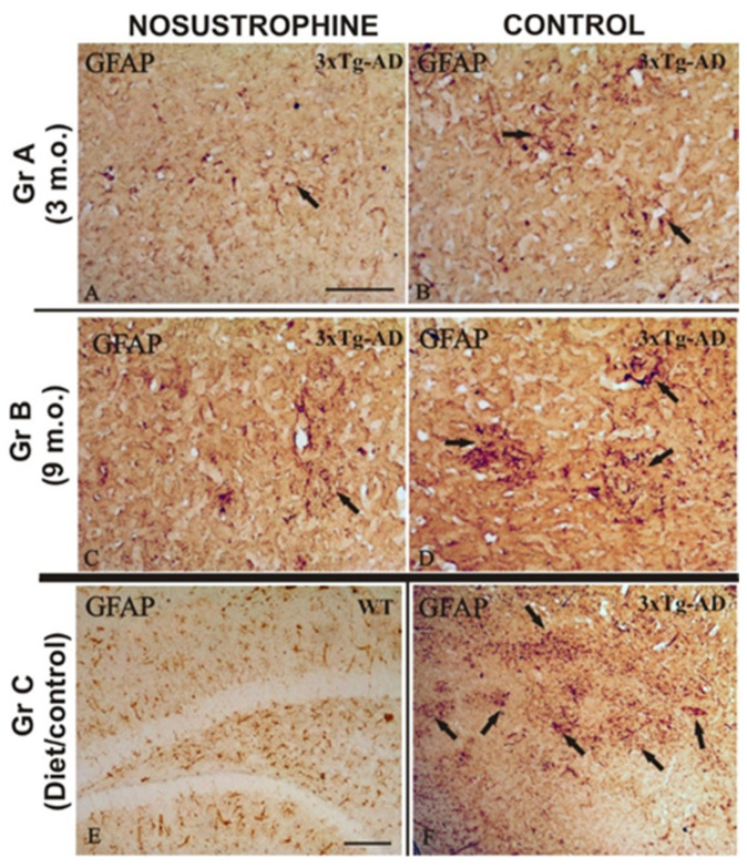
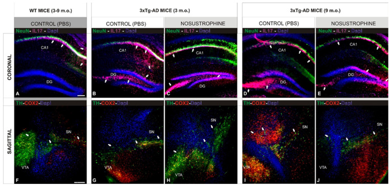
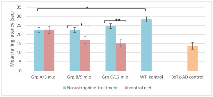
Neurodegeneration, characterized by the progressive deterioration of neurons and glial cells, is a feature of Alzheimer's disease (AD). The present study aims to demonstrate that the onset and early progression of neurodegenerative processes in transgenic mice models of AD can be delayed by a cocktail of neurotrophic factors and derived peptides named Nosustrophine, a nootropic supplement made by a peptide complex extracted from the young porcine brain, ensuring neuroprotection and improving neuro-functional recovery. Experimental 3xTg-APP/Bin1/COPS5 transgenic mice models of AD were treated with Nosustrophine at two different early ages, and their neuropathological hallmark and behavior response were analyzed. Results showed that Nosustrophine increased the activity of the immune system and reduced pathological changes in the hippocampus and cortex by halting the development of amyloid plaques, mainly seen in mice of 3-4 months of age, indicating that its effect is more preventive than therapeutic. Taken together, the results indicate the potent neuroprotective activity of Nosustrophine and its stimulating effects on neuronal plasticity. This study shows for the first time an effective therapy using nootropic supplements against degenerative diseases, although further investigation is needed to understand their molecular pathways.

Keywords: 3xTg-AD; Alzheimer’s disease; neuroprotection; nosustrophine; porcine brain extract.


Conflict of interest statement

The authors declare no conflict of interest.

Figures

Figure 1
Figure 1
Biochemical regulation of Nosustrophine in 3xTg-AD mice serum. Wild-type (WT) and AD-mice were treated with Nosustrophine. Most of the parameters analyzed show a significant correlation between an improved biochemical neuroprotection and mice treated with Nosustrophine, mainly in the early stages. (A,B): Inflammation response: Nosustrophine decreased C reactive protein (CRP) in young AD-mice. (CE): Antioxidant status improves with Nosustrophine: TAS increased in adult AD-mice (non-significant for WT) and GR increased mainly in WT and young AD-mice. (F): In lipid peroxidation (MDA), as marker of oxidative stress, no significant results were found. (G,H): Vitamin B6 levels increased in WT mice and Folate levels were higher in young AD-mice, both treated with Nosustrophine. (IK): Nosustrophine tends to decrease creatinine concentration and transaminases activity (GOT and GPT) in all groups being only significant for creatinine in WT mice. Significant values are represented as indicated * (p < 0.05), ** (p < 0.01) and *** (p < 0.001); Error bars: 95% CI. Data are expressed as mean ± standard error of the mean (SEM).
Figure 2
Figure 2
Progressive regulation of Aβ deposition in 3xTg-AD mouse brains treated with Nosustrophine. Comparative photomicrographs showing representative cortical and hippocampal brain sections of 3–9-month-old EB/3xTg-AD mice immunostained for Aβ. Images are presented according to the age group. (AD) Images of mice given Nosustrophine show early A-immunoreactive plaques in the cortical and hippocampus regions, whereas control sections display multiple A-immunoreactive plaques with a compact morphology at various stages; (E) nosustrophine-treated wild-type mice’s control brain slice, demonstrating no negative effects; (F) cortical slices of the control brain of EB/3Tg-AD transgenic mice are heavily marked by A-immunoreactive plaques that have a large diameter and a compact central core. Scale bar: 100 μm.
Figure 3
Figure 3
Nosustrophine modulates cortical density of β-amyloid plaques in early stages of AD mouse models. Comparative photomicrographs showing representative 3- and 9-month-old EB/3xTg-AD mouse brain sections stained by immunofluorescence antibody against β-amyloid. (A,C) Images showing the progressive accumulation of β-amyloid immunoreactive plaques in cortical regions of EB/3xTg-AD transgenic mice with no treatment (PBS); (B,D) in contrast, EB/3xTg-AD transgenic mice treated with Nosustrophine show a lower density of β-amyloid immunoreactive plaques in the early and middle stages of brain development. For abbreviations, see list. Scale bar: 100 μm.
Figure 4
Figure 4
Modulation of reactive immune system in AD mouse brains treated with Nosustrophine. Comparative photomicrographs showing representative brain regions of 3–9-month-old 3xTg-AD mouse brain sections immunostained for the lymphocyte marker CD11b. Images are presented according to the age group. (AD) Images are shown based on the age group. Images of the interior neocortical and hippocampus layers of 3xTg-AD transgenic mice depicting the increasing accumulation of CD11b-immunoreactive cells as an inflammatory response. In contrast, Nosustrophine-treated 3xTg-AD transgenic mice exhibit a low density of CD11b-immunoreactive cells at early and late phases of brain development; (E) nosustrophine-treated wild-type mice’s control brain slice, demonstrating no negative effects; (F) anatomical image of the control brain of 3xTg-AD transgenic mice demonstrating the abundance of inflammatory CD11b-immunoreactive cells in the damaged brain regions. Scale bar: 100 μm.
Figure 5
Figure 5
Nosustrophine regulate astrogliosis in AD mouse brains. Comparative photomicrographs showing representative brain regions of 4–9-month-old EB/3xTg-AD mice brain sections immunostained for GFAP. Images are presented according to the age group. (AD) Images showing the progressive accumulation of dystrophic reactive astrocytes from the dentate gyrus to the external layers of the neocortex. Note the contrast in astrocytosis density between Nosustrophine-treated mice and controls, where numerous immunoreactive GFAP clusters are observed; (E) control brain section of wild-type mice treated with Nosustrophine, showing no adverse effects; (F) control brain section of EB/3xTg-AD transgenic mice showing numerous inflammatory GFAP-immunoreactive cell clusters in the cortical and hippocampal brain areas. Scale bar: 100 μm.
Figure 6
Figure 6
Nosustrophine effect on neuropathological hallmarks in AD mouse models. Comparative photomicrographs showing representative 3 and 9-month-old EB/3xTg-AD mouse brain sections, early and middle stages of brain development, stained by antibodies against NeuN (neuronal nuclei), Cox2 (apoptosis), IL17 (proinflammatory cytokines), and TH (catecholaminergic neurons). (AE) Images of hippocampal regions showing the progressive accumulation of AD pathological markers in control (not treated) mice; (B,D) when compared with wild-type mice (A) and 3xTg-Ad treated with Nosustrophine (C,E); (FJ) Images of midbrain regions showing the massive accumulation of AD pathological markers in control (not treated) mice; (G,I) when compared with wild-type mice (F) and 3xTg-Ad treated with Nosustrophine (H,J). Scale bar: 100 μm.
Figure 7
Figure 7
Nosustrophine improves motor coordination in AD mouse models. Rotarod results of motor coordination and balance test of EB/3xTg-AD transgenic mice during four weeks. Notice the progressive decrease in rotarod performance that is directly related to the age of the mice. Latency to fall on the rotarod apparatus was significantly improved in mice treated with Nosustrophine, mainly in later stages as groups B (* p < 0.05) and C (** p < 0.01). Significant values represent means of latency of all mice in each group/trial and are expressed as standard error of the mean (±SEM).

Similar articles

Cited by

References

    1. Liu Y., Cheng F., Takeda S., Lai K., Lin L., Sakata R. Effects of porcine brain hydrolysate on impairment of cognitive learning ability in amyloid β(1-40)-infused rats. Anim. Sci. J. 2019;90:271–279. doi: 10.1111/asj.13054. - DOI - PubMed
    1. Tuszynski M., Yang J., Barba D., Hoi-Sang U., Bakay R., Pay M., Masliah E., Conner J., Kobalka P., Roy S., et al. Nerve Growth Factor Gene Therapy: Activation of Neuronal Responses in Alzheimer Disease. JAMA Neurol. 2015;72:1139–1147. doi: 10.1001/jamaneurol.2015.1807. - DOI - PMC - PubMed
    1. Froestl W., Muhs A., Pfeifer A. Cognitive enhancers (Nootropics). Part 1: Drugs interacting with receptors. J. Alzheimer’s Dis. 2014;41:961–1019. doi: 10.3233/JAD-140228. - DOI - PubMed
    1. Binder D., Scharfman H. Brain-derived neurotrophic factor. Growth Factors. 2004;22:123–131. doi: 10.1080/08977190410001723308. - DOI - PMC - PubMed
    1. Zhang L., Fang Y., Lian Y., Chen Y., Wu T., Zheng Y., Zong H., Sun L., Zhang R., Wang Z., et al. Brain-Derived Neurotrophic Factor Ameliorates Learning Deficits in a Rat Model of Alzheimer’s Disease Induced by Aβ1-42. PLoS ONE. 2015;10:e0122415. doi: 10.1371/journal.pone.0122415. - DOI - PMC - PubMed