. 2023 Aug 31;15(9):2261.
doi: 10.3390/pharmaceutics15092261.

Biomimetic Electrospun Self-Assembling Peptide Scaffolds for Neural Stem Cell Transplantation in Neural Tissue Engineering

Affiliations

Biomimetic Electrospun Self-Assembling Peptide Scaffolds for Neural Stem Cell Transplantation in Neural Tissue Engineering

Mahdi Forouharshad et al. Pharmaceutics. .

Abstract

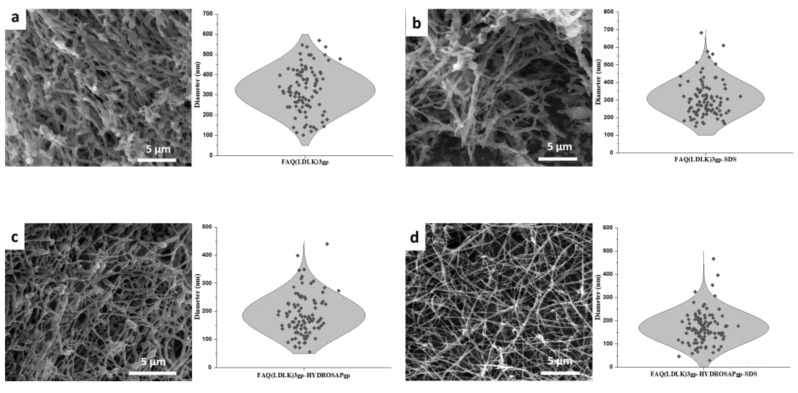
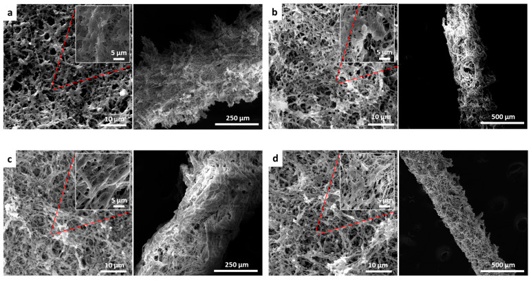
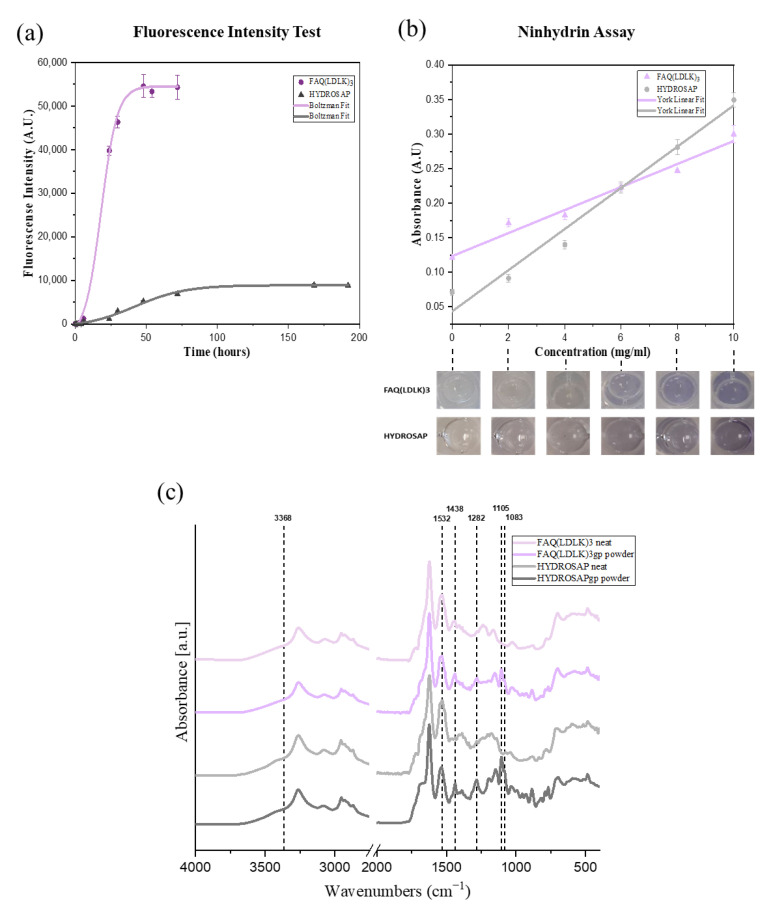
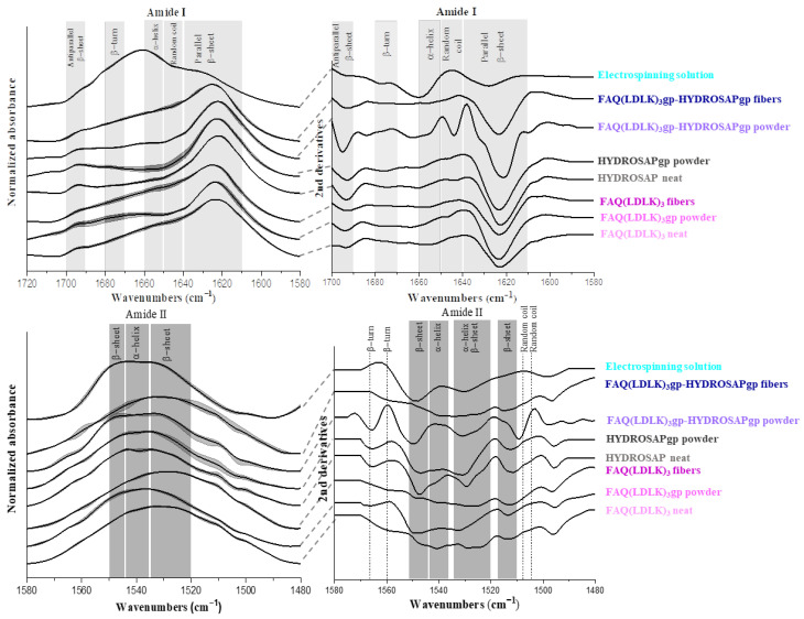
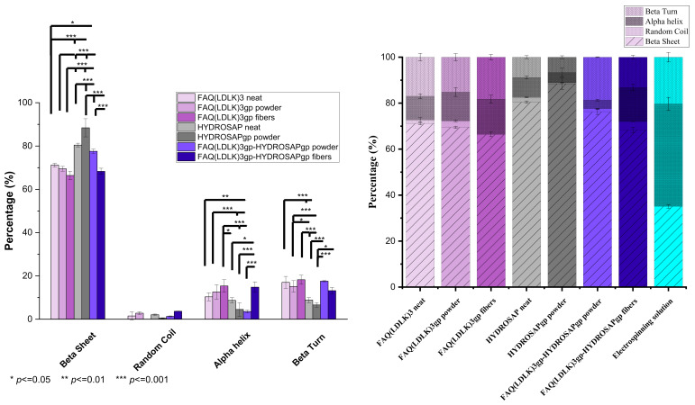
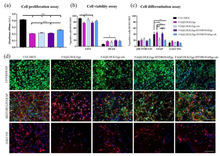
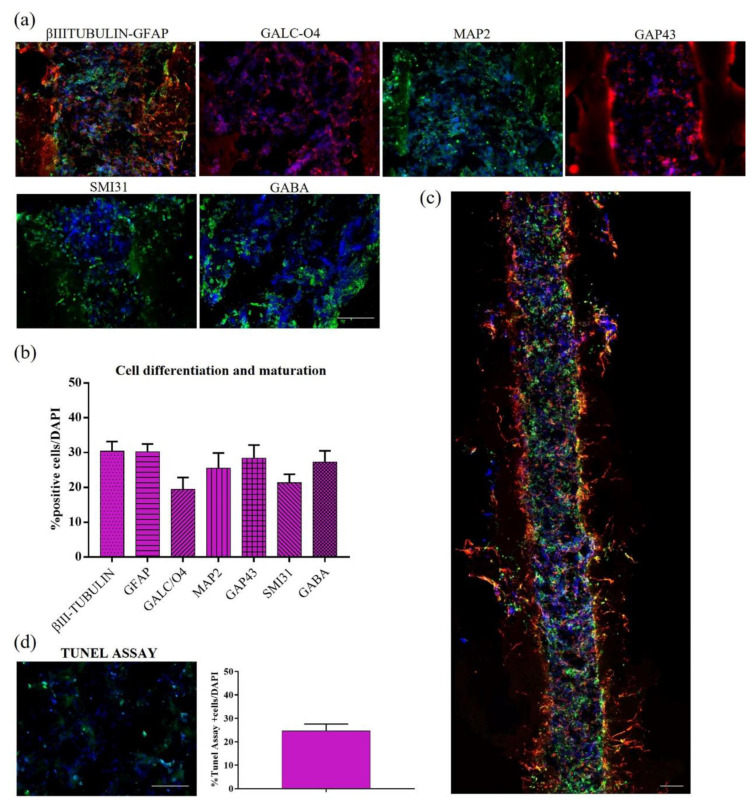
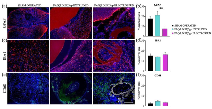
Spinal cord regeneration using stem cell transplantation is a promising strategy for regenerative therapy. Stem cells transplanted onto scaffolds that can mimic natural extracellular matrix (ECM) have the potential to significantly improve outcomes. In this study, we strived to develop a cell carrier by culturing neural stem cells (NSCs) onto electrospun 2D and 3D constructs made up of specific crosslinked functionalized self-assembling peptides (SAPs) featuring enhanced biomimetic and biomechanical properties. Morphology, architecture, and secondary structures of electrospun scaffolds in the solid-state and electrospinning solution were studied step by step. Morphological studies showed the benefit of mixed peptides and surfactants as additives to form thinner, uniform, and defect-free fibers. It has been observed that β-sheet conformation as evidence of self-assembling has been predominant throughout the process except for the electrospinning solution. In vitro NSCs seeded on electrospun SAP scaffolds in 2D and 3D conditions displayed desirable proliferation, viability, and differentiation in comparison to the gold standard. In vivo biocompatibility assay confirmed the permissibility of implanted fibrous channels by foreign body reaction. The results of this study demonstrated that fibrous 2D/3D electrospun SAP scaffolds, when shaped as micro-channels, can be suitable to support NSC transplantation for regeneration following spinal cord injury.

Keywords: 2D/3D scaffolds; electrospinning; regenerative medicine; secondary structures; self-assembling peptides; spinal cord injury.


Conflict of interest statement

The authors declare no conflict of interest.

Figures

Figure 1
Figure 1
SEM images of electrospun 2D scaffolds w/o post-treatment include fibers diameter distribution: (a) electrospun FAQ(LDLK)3gp with diameter distribution between 100 to 600 nm and an average diameter of 324.8 ± 11.1, (b) electrospun FAQ(LDLK)3gp-sds shows narrower diameter distribution with lower average diameter 307.9 ± 10.3 nm, (c) electrospun FAQ(LDLK)3gp-HYDROSAPgp shows diameter distribution between 80–400 nm with an average diameter of 185.3 ± 7.3 nm, and eventually (d) FAQ(LDLK)3gp-HYDROSAPgp-sds, comprising surfactant and mixed peptides, shows very narrow diameter distribution between 50–300 nm with an average diameter of 169.8 ± 7.0 nm.
Figure 2
Figure 2
SEM images of electrospun 2D and 3D scaffolds after post-treatment (annealing and crosslinking): (a) electrospun FAQ(LDLK)3gp lamina and channel show rough surface with fibers connection, (b) electrospun FAQ(LDLK)3gp-sds lamina and channel with more uniformity of fibers and surface after adding a surfactant, (c) electrospun FAQ(LDLK)3gp-HYDROSAPgp lamina and channel comprise defect-free fibers and even surface, (d) electrospun FAQ(LDLK)3gp-HYDROSAPgp-sds lamina and channel reveals very uniform fibers and surface after adding SDS and HYDROSAP (left: 2D lamina, right: 3D microchannels).
Figure 3
Figure 3
The crosslinking reaction progress was assessed using (a) Fluorescence intensity test by tracking the fluorescence intensity of blue fluorescent pigmentation generated by Genipin reaction with primary amines in peptides at 630 nm at different time points, (b) Ninhydrin assay by measuring the loss of free amine groups, reacted with ninhydrin, based on optical absorbance measurements at 570 nm, and (c) FTIR analysis to delineate the chemical and secondary structure changes in peptides after crosslinking by Genipin. The spectra were averaged and processed with the OriginPro software using Boltzmann fitting and York linear fitting.
Figure 4
Figure 4
FTIR spectra along with 2nd derivatives in amide I and II absorption region of entire samples: amide I (Up), Amide II (Down). Concerning amide I mode, the entire spectra, except the electrospinning solution, demonstrate the prominent absorption peaks in the region of β-sheet centered ~1630 cm−1. Electrospinning solution spectra show the main peak located in the region of α-helix. the spectral region corresponding to the amide II shows a broad peak in the region 1520–1545 cm−1 assigned to β-sheet and α-helix. Wavenumber ranges of the principal bands characteristic of peptide secondary structure [45,46].
Figure 5
Figure 5
Secondary structure content measured by three mathematical resolution enhancement methods of Fourier self-deconvolution, second derivative analysis, and band curve-fitting. Data are represented as average ±SEM (N = 3). Statistical analysis: One-way ANOVA followed by Tukey multiple comparison test. Statistical analysis shows significant differences between conditions (* p ≤ 0.05, ** p ≤ 0.01, and *** p ≤ 0.001) (Left), and the bar chart represents percentages of secondary structures as determined by the method of curve-fitting and peak deconvolution (Right).
Figure 6
Figure 6
Proliferation, viability, and differentiation assays of hNSCs seeded on 2D scaffolds of FAQ(LDLK)3gp, FAQ(LDLK)3gp-sds, FAQ(LDLK)3gp-HYDROSAPgp and FAQ(LDLK)3gp-HYDROSAPgp-sds after 7 days in vitro. (a) Colorimetric MTS assay for cell proliferation assessment. (b) LIVE/DEAD Cell Viability/Cytotoxicity test to determine cell viability. (c) Immunostainings for βIII-Tubulin (neurons in green), GFAP (astrocytes in red), and GALC/O4 (oligodendrocytes in red) markers. (d) Representative fluorescence images for cell viability assay (top), neural and astroglial differentiation (middle), and oligodendroglial differentiation (bottom). Live cells are labeled in green, and dead cells in red. Cell nuclei were stained with HOECHST (in blue). Data are represented as mean ± SEM. Statistical analysis shows significant differences between conditions (* p < 0.05; ** p ≤ 0.01; *** p < 0.001). All measures were performed in triplicate. Scale bar, 100 µm.
Figure 7
Figure 7
Cell differentiation and maturation of hNSCs seeded in HYDROSAP hydrogel and injected into the FAQ(LDLK)3gp microchannels (2 weeks in vitro). (a) Longitudinal sections of microchannels seeded with hNSCs. Neurons are labeled in green with βIIITubulin marker and astrocytes in red with GFAP; oligodendrocytes are stained with GALC/O4 in red, mature neurons in green with MAP2, growth-cones associated protein GAP43 in red, phosphorylated neurofilaments with SMI31 in green and GABAergic neurons in green. (b) Quantitative evaluation of neural markers for cell differentiation and maturation: βIII-Tubulin, GFAP, GALC-O4, MAP2, GAP43, SMI31, and GABA. (c) Full longitudinal section of a microchannel seeded with differentiated hNSCs progeny (βIII-Tubulin and GFAP stainings). (d) Tunel assay for apoptotic cells (green) inside the microchannels and their quantification. Cell nuclei are stained in blue with HOECHST. Data are represented as mean ± SEM (n = 8). Scale bars, 100 µm.
Figure 8
Figure 8
In vivo tests conducted to assess tissue response in sham-operated animals and those receiving FAQ(LDLK)3gp extruded and FAQ(LDLK)3gp electrospun scaffolds. Immunofluorescence staining: (a,b) GFAP, (c,d) IBA1, and (e,f) CD68 markers. Cell nuclei are made visible using DAPI staining (Blue). The reactive areas of GFAP+ cells (shown in red) detected near the implantation sites showed a significant difference between the FAQ(LDLK)3gp extruded and FAQ(LDLK)3gp electrospun experimental groups (** p ≤ 0.01). In the case of IBA1 and CD68 markers, the results on their reactivity areas showed no statistical differences among the experimental groups. Scale bar, 100 μm.

References

    1. Wang T., Nanda S.S., Papaefthymiou G.C., Yi D.K. Mechanophysical Cues in Extracellular Matrix Regulation of Cell Behavior. ChemBioChem. 2020;21:1254–1264. doi: 10.1002/cbic.201900686. - DOI - PubMed
    1. Kusindarta D.L., Wihadmadyatami H. Tissue Regeneration. IntechOpen; London, UK: 2018. The role of extracellular matrix in tissue regeneration. - DOI
    1. Onuwaje I., Phillips J.B. Handbook of Innovations in Central Nervous System Regenerative Medicine. Elsevier; Amsterdam, The Netherlands: 2020. Three-dimensional culture systems in central nervous system research; pp. 571–601.
    1. Kharbikar B.N., Mohindra P., Desai T.A. Biomaterials to enhance stem cell transplantation. Cell Stem Cell. 2022;29:692–721. doi: 10.1016/j.stem.2022.04.002. - DOI - PMC - PubMed
    1. de Freria C.M., Van Niekerk E., Blesch A., Lu P. Neural Stem Cells: Promoting Axonal Regeneration and Spinal Cord Connectivity. Cells. 2021;10:3296. doi: 10.3390/cells10123296. - DOI - PMC - PubMed